
Human Tenocytes
Cat.No.: CSC-C1584
Species: Human
Source: Tendon
Cell Type: Tenocyte
- Specification
- Background
- Scientific Data
- Q & A
- Customer Review
The vast majority of cells present in tendon are tenocytes (90-95%), which are specialized tendon fibroblasts responsible for extracellular matrix deposition. Tenocytes are embedded in a three-dimensional network of numerous extracellular matrix components, primarily composed of type I collagen fibrils (which account for more than 95% of tendon collagen), as well as other types of collagens (type III and type V), proteoglycans, elastin and fibronectin. These tendon-specific matrix components give tendon its resilience and biomechanical stability.
Tenocytes are an ideal model for in vitro studies of tendon regeneration and repair, the effects of cytokines and growth factors on tendon, and specific gene regulation and pathophysiological processes in tendon disorders. They are also useful tools for establishing in vitro disease models for high throughput and high content screening.
Human tenocytes provided by Creative Bioarray are isolated from healthy human patellar tendon. These tenocytes are cryopreserved at passage one and delivered frozen. Each vial of Human tenocytes contains more than 500,000 viable cells. Creative Bioarray also offers optimized media and reagents for the growth of human tenocytes, which are quality tested together and guaranteed to give optimal performance as a global solution for in vitro tenocyte culture.
Hyaluronic Acid Alleviates Oxidative Stress and Apoptosis in Human Tenocytes via Caspase 3 and 7
Rotator cuff tendinopathy (RCT) is the primary reason for shoulder surgery and its clinical management is still challenging. Hyaluronic acid (HA) has been shown to have anti-inflammatory effects in vitro and in vivo under RCT conditions, characterized by an exaggerated oxidative stress (OS). With these aims, a cell model of RCT was established by exposing primary human tenocytes to H2O2 for up to 72 h. Four different HAs by molecular weight were administered to measure nitric oxide (NO) and OS, apoptosis, and collagen 1 expression. The present study highlights that HAs characterized by a low molecular weight are able to counteract the H2O2-induced OS by decreasing the percentage of apoptotic cells and reversing the activation of caspase 3 and 7.
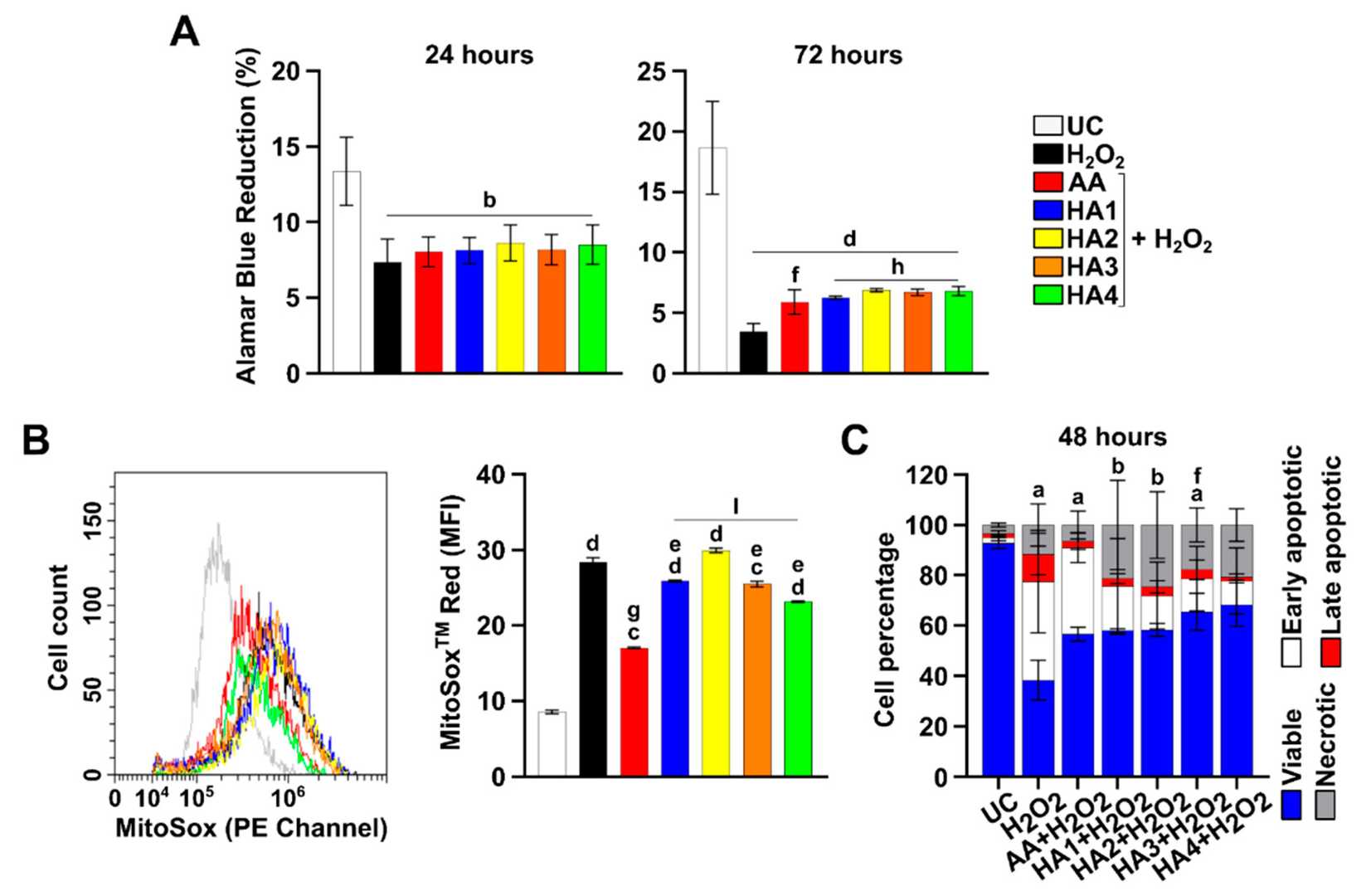
 Fig. 1. Cell proliferation, generation of superoxide anions, apoptosis, and caspase 3/7 activity in tenocytes exposed to hyaluronic acids under OS conditions (Gallorini, Marialucia, et al. 2022).
Untreated control (UC); hydrogen peroxide 2 mM (H2O2); ascorbic acid μg/mL (AA); Artrosulfur HA® (HA1), Synolis VA® (HA2), Hyalubrix® (HA3) and Hyalgan® (HA4) at a final concentration of 1 mg/mL.
Dehydrated Human Amniotic Membrane Regulates Tenocyte Expression and Angiogenesis In Vitro
This study evaluated the effects of micronized dehydrated human amnion/chorion membrane (μdHACM) on the inflammatory environment and hypervascularity associated with tendinopathy. Stimulation of human tenocytes with interleukin-1 beta (IL1β) induced the expression of inflammatory and catabolic markers, resulting in secretion of active MMPs and type 3 collagen that is associated with a degenerative phenotype. Treatment with μdHACM diminished the effects of IL1β, reducing the expression of inflammatory genes, proteases, and extracellular matrix components, and decreasing the presence of active MMP and type 3 collagen. Additionally, a co-culture model was developed to evaluate the effects of μdHACM on angiogenesis associated with tendinopathy.
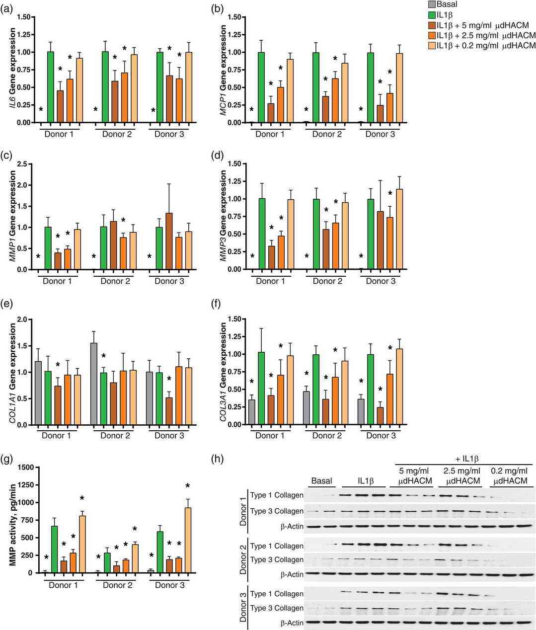 Fig. 2. Treatment with μdHACM reduces IL1β induced changes in tenocytes. (Moreno, Sarah E., Michelle Massee, and Thomas J. Koob. 2022)
Ask a Question
Write your own review
- You May Also Need
Description: HNPC from Creative Bioarray are isolated from nucleus pulposus of human intervertibral disc. HNPC are cryopreserved at primary or passage one culture and delivered frozen. Each vial contains >5 x ...

Description: HS from Creative Bioarray Research Laboratories are isolated from human synovium. HS are cryopreserved at passage one cultures and delivered frozen. Each vial contains >5 x 10^5 cells in 1 ml ...
Description: HC-a from Creative Bioarray are isolated from human articular cartilage. HC-a are cryopreserved at primary cultures and delivered frozen. Each vial contains >5 x 10^5 cells in 1 ml volume. HC-a are ...
Description: Description: Human skeletal muscle cells are derived from whole muscle that has been dissociated into single cells and frozen. These muscle cells are from a single donor. They enable researchers to ...
Description: HAFC from Creative Bioarray are isolated from annulus fibrosus of human intervertibral disc. HAFC are cryopreserved at primary or passage one culture and delivered frozen. Each vial contains >5 x ...

Description: RFP Expressing Human Chondrocytes provided by Creative Bioarray are puromycin-selected from primary human chondrocytes infected with lentivirus expressing RFP. RFP Expressing Human Chondrocytes are ...