Porcine Retinal Pigment Epithelial Cells
Cat.No.: CSC-C4740Z
Species: Pig
Source: Retina; Eye
Cell Type: Epithelial Cell
- Specification
- Background
- Scientific Data
- Q & A
- Customer Review
Never can cryopreserved cells be kept at -20 °C
Porcine retinal pigment epithelial (RPE) cells are epithelial cells isolated from the retinal pigment epithelium of pig (Sus scrofa) eyes. They are an extensively used in vitro model for ocular and retinal research due to the high similarity between porcine and human eyes in size, retinal structure, and physiological properties. Porcine RPE cells are often used as a translational model when human primary RPE cells are not available.
In morphology, porcine RPE cells share the same polygonal, cobblestone-like shape of other RPE and grow as tightly packed, adherent monolayers with robust cell-cell junctions. If maintained in the right conditions, porcine RPE cells exhibit apical-basal polarity and express common RPE markers (e.g., RPE65, cytokeratins, ZO-1, and bestrophin-1) to signify their epithelial differentiated phenotype. Porcine RPE cells are able to form functional tight junctions to maintain the outer blood-retinal barrier.
In function, porcine RPE cells maintain many of the physiological functions of native RPE including phagocytosis of photoreceptor outer segments, transport of ions and nutrients, secretion of growth factors, and involvement in the visual cycle. These cells are therefore utilized in a wide variety of research applications including studies of retinal development and retinal degeneration as well as disease models of age-related macular degeneration (AMD) and diabetic retinopathy. Porcine RPE cells are also commonly used to study oxidative stress and inflammation, as well as drug permeability across the retina.
Supplemented Media Leads to Significant ppRPE Loss
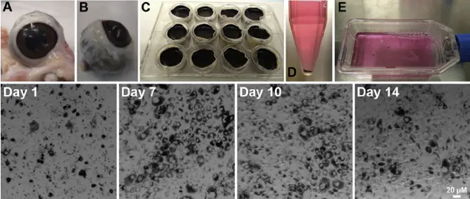
AMD is a prevalent cause of vision loss in the elderly. To better understand its pathogenesis, Wagner et al. developed an ex vivo model using ppRPE cells exposed to sodium iodate (NaIO3) for degeneration and supplemented media (BSA, HPR, ROOS) to promote retinal deposits. These cells were then co-cultured with porcine neuroretina explants.
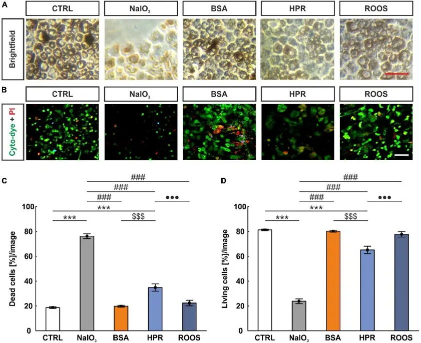
All five groups of ppRPE cells showed typical dark pigmentation and cobblestone morphology (Fig. 1A). After retinal supplemented media was applied, live and dead cell staining was performed (Fig. 1B). Compared to CTRL ppRPE cells, all supplemented groups had significantly fewer cells. The NaIO3 group had the lowest cell count. The NaIO3 group had the highest percentage of dead cells compared to other groups. The HPR group also had significantly more dead cells than CTRL, BSA, and ROOS groups (Fig. 1C). The NaIO3 group had the lowest percentage of living cells compared to other groups. The HPR group had significantly fewer living cells than CTRL, BSA, and ROOS groups (Fig. 1D).
GIBICO's Fetal Bovine Serum is not pre-aged. When stored at 2 to 8 °C, various proteins and lipoproteins (e.g., cold agglutinin, fibrinogen, hyaluronan, etc.) in the serum may aggregate and form a precipitate or visible turbidity. This should not affect the quality of the serum. It is recommended that fetal bovine serum be stored at -20 °C to avoid repeated freezing and thawing.
Ask a Question
Average Rating: 4.0 | 1 Scientist has reviewed this product
Effective preservation
Creative Bioarray's cell products are top notch. I had great results with the process and everything went smoothly.
02 Aug 2023
Ease of use
After sales services
Value for money
Write your own review
Description: Porcine Corneal Epithelial Cells from Creative Bioarray are isolated from corneal tissue of porcine. Porcine Corneal Epithelial Cells are grown in a T25 tissue culture flask pre-coated with ...
Description: Porcine Brain Vascular Fibroblasts from Creative Bioarray are isolated from brain tissue of porcine. Porcine Brain Vascular Fibroblasts are grown in T75 tissue culture flasks pre-coated with ...
Description: Porcine Thymus Endothelial Cells from Creative Bioarray are isolated from thymus tissue of porcine. Porcine Thymus Endothelial Cells are grown in T25 tissue culture flasks pre-coated with ...

Description: Porcine Kidney Endothelial Cells from Creative Bioarray are isolated from kidney tissue of porcine. Porcine Kidney Endothelial Cells are grown in T25 tissue culture flasks pre-coated with ...

Description: Pig bone marrow from Creative Bioarray is procured from the fresh femurs. Bone Marrow-CD34+ stem/progenitor cells are positively isolated using a direct immunomagnetic CD34 MicroBead labeling system.
Description: Porcine Primary Thymus Fibroblasts from Creative Bioarray are isolated from thymus tissue of porcine. Porcine Primary Thymus Fibroblasts are grown in T75 tissue culture flasks pre-coated with ...