Primary Human Melanocyte Cells
Cat.No.: CSC-C4370X
Species: Human
Source: Foreskin
Cell Type: Melanocyte
- Specification
- Background
- Scientific Data
- Q & A
- Customer Review
These cells were originated using CSCM 2.5 Complete Serum-Free Medium, are available at <12 Cumulative Population Doublings (CPD) in vitro [Passage 3]. This vial will initiate a Passage 4 cell culture in a 75cm2 flask.
These cells are available in both cryopreserved vials as well as in 25cm2 and 75cm2 proliferating culture flasks.
Each vial or flask of cells is shipped to Customer with Bac-O (antibiotic) and SuperEnergy™ (animal derived growth factors) or SuperEnergy-R™ (human recombinant growth factors) at no additional cost.
Primary Human Melanocyte Cells are progenitor cells extracted from the epidermal layer of human skin, responsible for the synthesis and distribution of melanin pigment. These are non-transformed cells that maintain the true physiological behavior of native melanocytes including the ability to form dendrites and respond to UV light and paracrine signals from keratinocytes, unlike immortalized lines.
In culture these cells display a bipolar or multipolar dendritic architecture and develop as adherent monolayers. Fibroblasts are cultivated in a specialized basal medium supplemented with growth factors such as basic Fibroblast Growth Factor (bFGF) and agents that increase intracellular cyclic AMP (e.g., phorbol esters or cholera toxin) to inhibit overgrowth and maintain phenotypic. Standard incubation is 37°C and 5% CO2.
These cells are an important model for the studies of melanogenesis, UV induced DNA damage response and skin pigmentation diseases, such as vitiligo and albinism. They are also utilized widely in the study of early events in the transformation of melanoma and in the evaluation of chemopreventive agents, providing a biologically meaningful alternative to fully converted cancer cell lines.
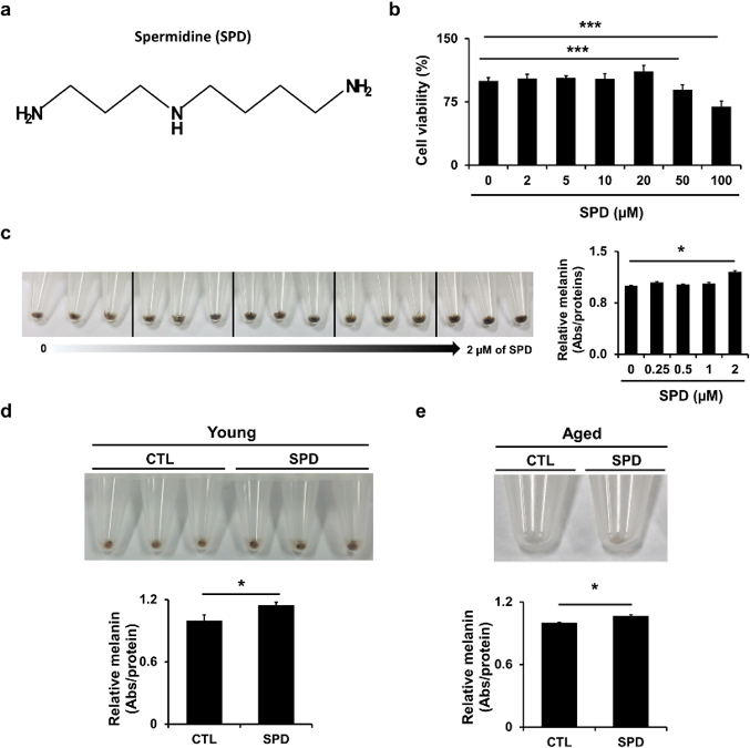
SPD Treatment Increases Melanin Production
Spermidine (SPD), a polyamine with known anti-aging properties, has an uncharacterized role in skin pigmentation. Given the lack of effective treatments for hypopigmentation disorders, Brito et al. investigated SPD's effects on melanogenesis in normal human primary melanocytes and the MNT-1 melanoma cell line.
Cytotoxicity assays confirmed that SPD concentrations up to 25 µM were safe for MNT-1 cells, while 50-100 µM reduced viability (Fig. 1b). Subsequent experiments used non-toxic doses. SPD treatment significantly increased melanin content in MNT-1 cells in a dose-dependent manner (Fig. 1c). Furthermore, SPD enhanced melanin production in both young and aged primary melanocytes (by 15 ± 5% and 12 ± 2%, respectively) (Fig. 1d, e). These findings identify SPD as a novel natural agent capable of stimulating pigmentation across different cell types and donor ages.
Ask a Question
Write your own review

Description: Microglia, one of the glial cell types in the CNS, is an important integral component of neuro-glial cell network. They have been observed in the brain parenchyma from the early stage of development ...
Description: Primary Human Synovial Microvascular Endothelial Cells were initiated by elutriation from dispase dissociated normal human synovial tissue .These cells were originated using Complete Serum-Free ...

Description: Creative Bioarray's Normal Human Dermal Microvascular Endothelial Cells (HD-MVECn), when grown in Creative Bioarray's LIVas Medium, provides an ideal culture model, with or without human VEGF, for ...
Description: Human Corneal Epithelial Cells from Creative Bioarray are isolated from normal human corneal tissue. Human Corneal Epithelial Cells are grown in T25 tissue culture flasks pre-coated with ...
Description: Adult normal Primary Sebocytes are a vital research tool for a variety of applications from acne, seborrhoea and other sebaceous gland-related diseases studies in skin biology/cosmetic programs. ...
Description: Normal primary adult melanocytes are a vital research tool for a variety of applications from malignant melanoma research to pigmentation studies as well as for cosmetics and skin biology programs. ...