
Human Dermal Microvascular Endothelial Cells-Neonatal (HD-MVECn)
Cat.No.: CSC-C4098X
Species: Human
Source: Dermis; Skin
Cell Type: Endothelial Cell; Microvascular Cell
- Specification
- Background
- Scientific Data
- Q & A
- Customer Review
Cell Features:
Human Bladder Epithelial Cells are isolated from Human Bladder Epithelial tissue and are cyropreserved as secondary cells.
Human Bladder Smooth Muscle Cells are isolated from Human Bladder Smooth Muscle tissue and are cryopreserved as secondary cells.
Human Bladder Fibroblast Cells are isolated from Human Bladder tissue and are cryopreserved as secondary cells.
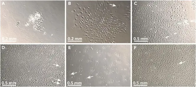
Human Dermal Microvascular Endothelial Cells-Neonatal (HD-MVECn) are primary endothelial cells isolated from the microvascular endothelium of neonatal human dermis, including the capillaries, arterioles, venules, and lymphatic microvessels. The endothelium is the structural and functional basis of dermal microcirculation that is integral to skin homeostasis, including processes such as barrier and nutrient exchange function, angiogenesis, and inflammation. Neonatal-derived cells have increased proliferative potential and a more stable endothelial phenotype than adult endothelial cells, and are well suited to physiologic and pathologic vascular biology studies.
Morphologically, HD-MVECn in culture are cobblestone in appearance and form closely adherent monolayers that are positive for key endothelial markers including VE-cadherin, CD31, vWF, and VEGFR-2. They also maintain a functional phenotype, including strong angiogenic potential, the ability to internalize Dil-Ac-LDL, and the capacity to form tubes in extracellular matrix matrices. These cells respond dynamically to cytokines and inflammatory stimuli, upregulating adhesion molecules (ICAM-1, VCAM-1, E-selectin) that allow modeling of leukocyte adhesion, endothelial activation, and microvascular permeability. HD-MVECn are used widely in studies of angiogenesis, skin inflammation, wound healing, microvascular remodeling, and endothelial barrier biology, with utility in drug screening and dermal toxicity testing, transdermal delivery, and tissue-engineering contexts such as 3D vascularized skin constructs and skin-on-chip models. In general, HD-MVECn represent a physiologically relevant model for studying human microvascular endothelial biology.
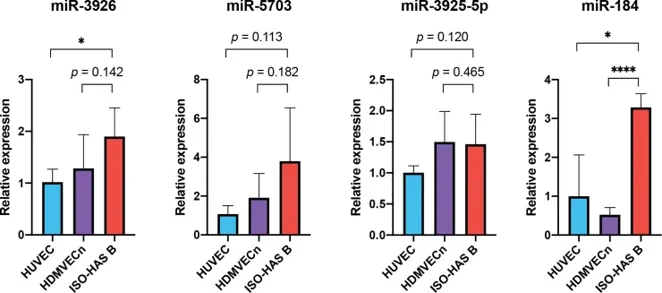
Serum Expression Levels of EV-Derived miR-3926, miR-5703, miR-3925-5p and miR-184 Showed a Correlation with the Disease Progress in a Angiosarcoma Patient
Cutaneous angiosarcoma is a rare and aggressive malignancy of the skin. In this study, Yokoi et al. seek to identify an alteration of serum-derived extracellular vesicle (EV) in angiosarcoma (AS) patients and to evaluate the clinical applicability as a novel circulating biomarker.
To identify miRNAs differentially expressed between AS patients and healthy controls (HCs), miRNA microarray analysis was performed with serum EV-miRNA samples isolated from 15 AS patients and 5 HCs. The results of the microarray analysis showed 73 and 100 miRNAs were significantly upregulated and downregulated, respectively, in AS patients relative to HCs. To explore if AS cells could be the source of these miRNAs, a TaqMan MicroRNA Assay was performed on an AS cell line (ISO-HAS B) and two kinds of endothelial cell, human umbilical vein endothelial cells (HUVEC) and human dermal microvascular endothelial cells neonatal (HDMVECn). The results demonstrated an increased expression level of miR-3926, miR-5703, miR-3925-5p and miR-184 in ISO-HAS B relative to HUVEC and an increased expression level of miR-3926, miR-5703 and miR-184 in ISO-HAS B relative to HDMVECn (Fig. 1).
Ask a Question
Write your own review
Description: Microglia, one of the glial cell types in the CNS, is an important integral component of neuro-glial cell network. They have been observed in the brain parenchyma from the early stage of development ...

Description: Primary Human Synovial Microvascular Endothelial Cells were initiated by elutriation from dispase dissociated normal human synovial tissue .These cells were originated using Complete Serum-Free ...
Description: Human Corneal Epithelial Cells from Creative Bioarray are isolated from normal human corneal tissue. Human Corneal Epithelial Cells are grown in T25 tissue culture flasks pre-coated with ...
Description: Adult normal Primary Sebocytes are a vital research tool for a variety of applications from acne, seborrhoea and other sebaceous gland-related diseases studies in skin biology/cosmetic programs. ...

Description: Normal primary adult melanocytes are a vital research tool for a variety of applications from malignant melanoma research to pigmentation studies as well as for cosmetics and skin biology programs. ...

Description: Primary Human Vascular Smooth Muscle Cells are pooled primary isolates from 250 individual human neonatal donor umbilical arteries.These cells were originated using CSCM Complete Medium, are ...