
Rat Cardiomyocytes
Cat.No.: CSC-C1151Z
Species: Rat
Source: Heart
Cell Type: Cardiomyocyte
- Specification
- Background
- Scientific Data
- Q & A
- Customer Review
Upon receipt, if no dry ice is left in the package, thaw and use cells immediately.
If there is dry ice left in the package, store it in liquid nitrogen immediately upon arrival.
When stored at the recommended storag
Rat cardiomyocytes are terminally differentiated, specialized muscle cells isolated from rat heart tissue. Two principal preparations are utilized in research: neonatal rat cardiomyocytes (NRCMs) , isolated from 1-2 day-old pups, and adult rat ventricular myocytes (ARVMs), harvested from mature animals. NRCMs exhibit a characteristic elongated, striated morphology and display spontaneous, rhythmic contractions in culture, while ARVMs retain a rod-shaped morphology with organized sarcomeres and functional excitation-contraction coupling.
A key advantage of rat cardiomyocytes is their high physiological relevance. Isolated cells closely resemble the intact myocardium in many respects, preserving critical in vivo features such as sarcomeric alignment, ion channel expression, and calcium-handling properties. This makes them superior to immortalized cell lines for studying fundamental cardiac biology. Neonatal cells are particularly advantageous due to their relative ease of isolation, robust spontaneous beating, and high viability in culture. They express well-defined electrophysiological properties including L- and T-type calcium currents and distinct action potentials. Adult cells, while more technically challenging to isolate and maintain, provide a model that more faithfully represents mature myocardial function.
Rat cardiomyocytes serve as an invaluable platform for investigating cardiac development, hypertrophy, ischemia-reperfusion injury, and drug-induced cardiotoxicity. They are amenable to live-cell imaging, electrophysiological recording, and genetic manipulation via viral transduction. Despite inherent limitations-including species-specific differences in electrophysiology compared to humans and the post-mitotic nature of adult cells-rat cardiomyocytes remain a gold-standard ex vivo model bridging fundamental cardiovascular biology and preclinical drug discovery.
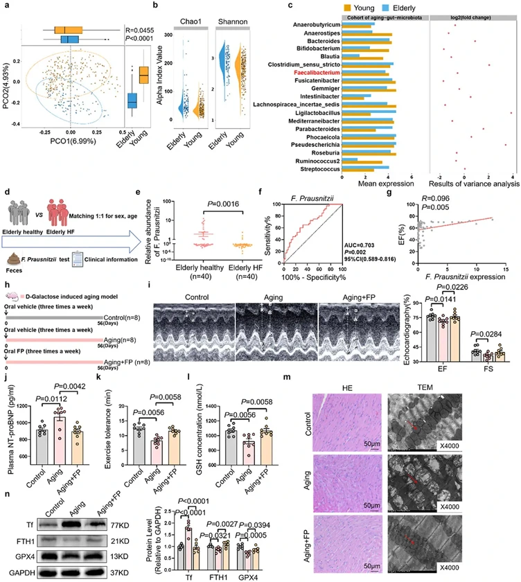
Faecalibacterium Prausnitzii Prevents Age-Related Heart Failure by Suppressing Ferroptosis in Cardiomyocytes
Aging is a primary driver of the escalating prevalence of heart failure (HF). Age-associated gut microbiota dysbiosis has been implicated in various age-related diseases, yet its role in age-related HF remains largely unexplored. In this study, we sought to explore the potential link between age-related gut microbiota alterations and HF in the elderly.
We analyzed a publicly available single-cell sequencing dataset, which revealed markedly increased ferroptosis activity in cardiac myocytes of elderly individuals compared to their younger counterparts. Notably, treatment with the ferroptosis inhibitor, ferrostatin-1, mitigated cardiac ferroptosis and prevented cardiac dysfunction in aging rats. Furthermore, fecal microbiota transplantation from elderly HF patients significantly increased cardiac ferroptosis activity and induced cardiac dysfunction in healthy recipient rats. Integrated 16S rRNA sequencing and PCR quantification revealed a marked depletion of Faecalibacterium prausnitzii (F. prausnitzii) in elderly individuals, with a more pronounced decline in elderly patients with HF. Oral administration of F. prausnitzii or its metabolite butyrate effectively attenuated age-related HF through inhibiting ferroptosis. Additionally, gene-editing techniques were employed to generate F. prausnitzii BCoAT mutant deficient in butyrate production. Intriguingly, the protective effect was lost in the butyrate-deficient F. prausnitzii strain.
These findings highlight the critical role of aged microbiota-induced ferroptosis in HF and propose F. prausnitzii or butyrate may serve as potential targets for the prevention and treatment of age-related HF.
Ask a Question
Write your own review
Description: The thoracic aorta is located in the chest cavity and gives off arteries that branch to the esophagus, pericardium, lungs, and trachea. The thoracic aorta can be subdivided into the ascending aorta, ...
Description: Rat Podocytes are isolated from normal rat kidney. The cells are characterized by immunofluorescence with antibodies specific to podocin, Ang1, Nephrin, ACTN4, NPHS2. T25 flasks is required for cell ...
Description: Rat Bronchial Smooth Muscle Cells are isolated from normal rat bronchi tissue. Rat Bronchial Smooth Muscle Cells are characterized by immunofluorescence with antibodies specific to alpha-actin. T25 ...
Description: Guinea Pig Endothelial Cells from Creative Bioarray are isolated from guinea pig tissue. Prior to shipping, cells at passage 2 are detached from flasks and immediately cryopreserved in vials. Each ...
Description: Rat Lung Epithelial Cells are isolated from normal rat lung tissue. The cells are characterized by immunofluorescence with antibodies specific to CK-18, CK-19. T25 flasks is required for cell ...
Description: Rat Vein Endothelial Cells from Creative Bioarray are isolated from inferior vena cava tissue of 6-8 week old laboratory Sprague-Dawley rat. Rat Vein Endothelial Cells are grown in T75 tissue culture ...