
Normal Human Epidermal Keratinocytes
Cat.No.: CSC-C4071X
Species: Human
Source: Epidermis; Skin
Cell Type: Keratinocyte
- Specification
- Background
- Scientific Data
- Q & A
- Customer Review
Cell Features:
HEKn are cryopreserved as primary cells. Cells are isolated from neonatal human foreskin and expanded once in culture vessels before cryopreservation.
HEKa are cryopreserved as primary cells. Cells are isolated from adult human skin and expanded once in culture vessels before cryopreservation.
HEK can be grown without serum, phenol red or antimicrobials when cultured in DermaLife medium.
HEK are extensively tested for quality and optimal performance.
Creative Bioarray guarantees performance and quality.
Normal human epidermal keratinocytes (NHEKs) are primary keratinocytes isolated from normal human skin, typically neonatal or adult epidermis. They retain basal markers p63 and cytokeratin 5/14 and, upon differentiation, express cytokeratin 1/10, involucrin, filaggrin, and loricrin. In low-calcium serum-free medium, NHEKs maintain a proliferative basal phenotype; calcium switch or air-liquid interface culture induces terminal differentiation, stratified epidermal architecture, cornified envelope formation, and barrier development. This enables functional studies of epidermal homeostasis, barrier integrity, innate immune signaling, and cutaneous inflammation.
Compared with immortalized keratinocyte lines such as HaCaT, NHEKs better preserve donor genetic background, native receptor expression, and physiological signaling. They are more human-relevant than animal keratinocytes for dermatological research. NHEKs are widely used to model wound healing, psoriasis, atopic dermatitis, contact dermatitis, photoaging, and skin carcinogenesis, and to evaluate drugs, cosmetics, irritants, sensitizers, and wound-care products. They are compatible with monolayer culture, 3D organotypic skin equivalents, and co-culture with fibroblasts, melanocytes, or immune cells.
Overall, NHEKs provide a robust, human-relevant, and physiologically characterized platform for cutaneous biology, disease modeling, safety assessment, and translational dermatological research.
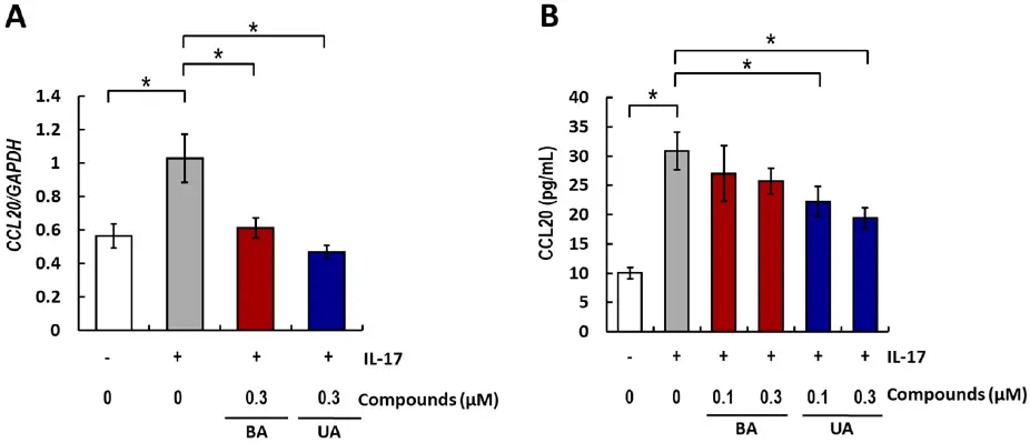
Effects of Betulinic Acid and Ursolic Acid on IL-17-Induced CCL20 Release in Normal Human Epidermal Keratinocytes
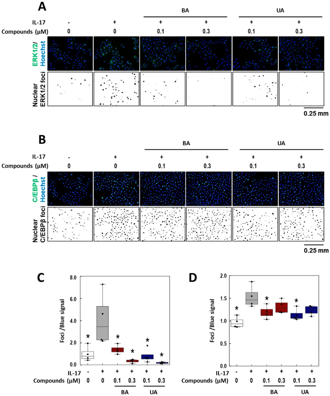
Psoriasis is a chronic inflammatory skin disease characterized by erythema, infiltration, and scaling, which is mainly caused by interleukin (IL)-17. The use of molecular targeted drugs in specific therapies offers high efficacy; however, high medical costs and a significant risk of side effects highlight the need for novel therapeutic agents. We previously observed that Morus alba extract (MAE) suppressed IL-17-induced CCL20 mRNA expression in normal human epidermal keratinocytes (NHEKs). In this study, we focused on the IL-17 signaling pathway and investigated the effects of pentacyclic triterpenoids, betulinic acid (BA), and ursolic acid (UA), which are present in MAE, on NHEK cells. Real-time reverse transcription polymerase chain reaction (RT-PCR) and enzyme-linked immunosorbent assay (ELISA) revealed that both BA and UA suppressed CCL20 expression, while only UA alone inhibited CCL20 release. ELISA using specific inhibitors demonstrated that both the p38 and extracellular-signal-regulated kinase 1/2 (ERK1/2) pathways were crucial for IL-17-induced CCL20 release in NHEK. UA effectively suppressed ERK1/2 nuclear localization and moderately affected p38 phosphorylation. These results indicated that UA is a potential seed compound for psoriasis treatment through its targeting of the IL-17 pathway.
Ask a Question
Write your own review
Description: Melanocytes are melanin-producing neural crest-derived cells located in the bottom layer (the stratum basale) of the skin's epidermis, the middle layer of the eye (the uvea), the inner ear, vaginal ...
Description: Creative Bioarray's normal Human Epidermal Melanocytes neonatal (HEMn), when grown in Creative Bioarray's LIDer M, provide an ideal low serum culture model, without PMA or Cholera toxin, for the ...
Description: The epithelial layer of the skin provides an essential function as a protective barrier against insult from the outside environment. The major cell type in this layer is keratinocytes which consists ...
Description: Neonatal Human Epidermal Keratinocytes (NHEK) are cultured by first, isolating the keratinocytes from the epidermis of the neonatal skin and then, establishing a primary keratinocyte culture. Upon ...
Description: The epithelial layer of the skin provides an essential function as a protective barrier against insult from the outside environment. The major cell type in this layer is keratinocytes which consists ...
Description: Creative Bioarray's normal Human Epidermal Melanocytes adult (HEMa), when grown in Creative Bioarray's LIDer Ma Medium, provide an ideal low serum culture model, without PMA or Cholera toxin, for the ...