Rat Macrophage
Cat.No.: CSC-C1801
Species: Rat
Source: Bone Marrow
Cell Type: Macrophage
- Specification
- Background
- Scientific Data
- Q & A
- Customer Review
RMa-bm are isolated from adult rat bone marrow tissue. Cells are harvested after purification and delivered frozen. Each vial contains >1 x 10^6 cells in 1 ml volume. RMa-bm is characterized by immunofluorescent method with antibody to CD 11b. RMa-bm is negative for mycoplasma, bacteria, yeast and fungi. RMa-bm is guaranteed to further culture in the condition provided by Creative Bioarray.
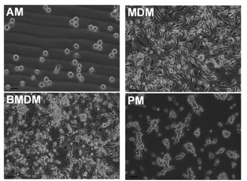
Rat macrophages are a diverse population of innate immune cells that are ubiquitous in rat tissues and have crucial functions in immunity, inflammation, tissue repair, and homeostasis. Rat macrophages can be derived from various anatomical sources, mainly peritoneum, lungs, bone marrow, and spleen and liver. Morphologically, resting cells are adherent with spindle-like or polygonal shapes and have eccentrically located kidneyshaped nuclei and abundant cytoplasm with lysosomes, whereas activated cells (e.g., by LPS) are irregular in shape, have condensed nuclei, and display prominent phagocytic granules in the cytoplasm. Primary macrophages are terminally differentiated and are adherent dependent with limited proliferation, whereas immortalized macrophage cell lines, such as NR8383, have an increased proliferative capacity (doubling time 24-36 h) and can be stably passaged.
Functionally, they are involved in phagocytosis of pathogens and apoptotic cells, production of pro-inflammatory (TNF-α, IL-1β) or anti-inflammatory cytokines (IL-10), and antigen presentation. Researchers extensively utilize rat macrophages for studies of inflammation, infection, tumor immunology and metabolic diseases as well as drug screening and toxicological assessments because these cells show high similarity to human macrophages and work well with disease models developed in rats.
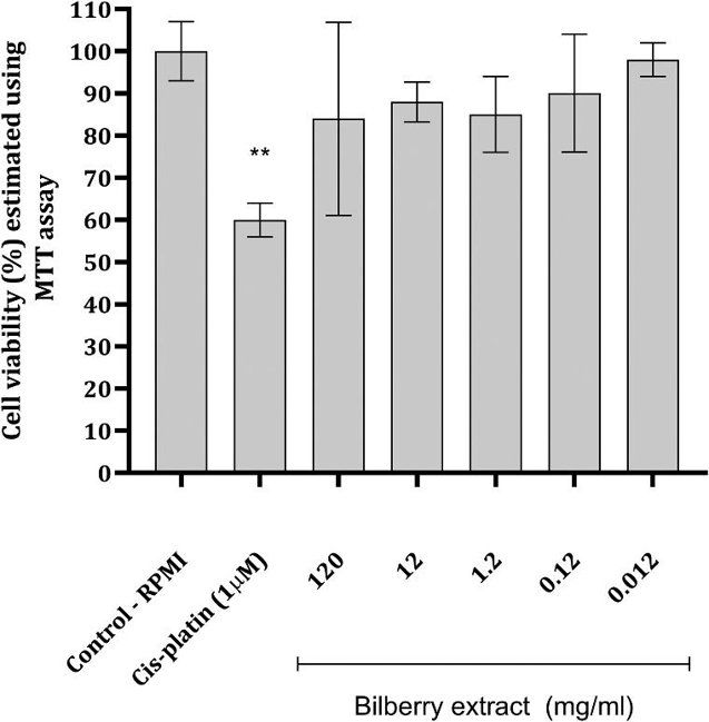
Effect of Bilberry Extract on Macrophage Viability and Myeloperoxidase Activity
In vitro experiments, animal studies, and clinical trials suggest that anthocyanin has antioxidant, anti-inflammatory, anti-diabetic, anti-hypertensive, and anti-dyslipidemic effects. Its health benefits are mainly due to its antioxidant properties. Stojanovic et al. assessed the anti-inflammatory potential of anthocyanin-rich bilberry extract by examining its antioxidant activity and effects on rat peritoneal macrophage inflammatory responses.
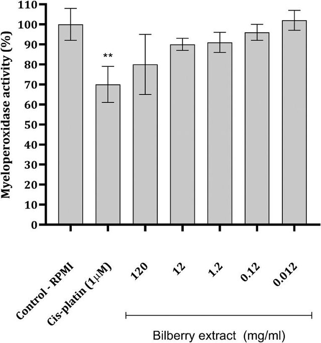
Peritoneal macrophages (Mφs) exposed to bilberry extract at different concentrations showed no significant toxicity in the MTT assay (Fig. 1). The highest concentration of bilberry extracts reduced Mφs viability by nearly 20%, while concentrations from 12 to 0.12 mg/mL caused a slight decrease of about 10%. The lowest concentration had no significant effect on cell viability (Fig. 1). In contrast, the standard drug cisplatin (CP) at 1 μM significantly reduced cell viability by around 40% (Fig. 1). Incubation of Mφs with bilberry extract did not significantly decrease myeloperoxidase (MPO) activity (Fig. 2). The most noticeable decrease in MPO activity, though non-significant, occurred with the highest extract concentration. However, CP significantly reduced MPO activity.
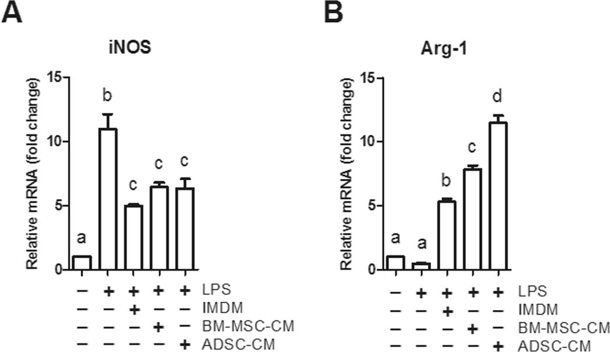
ADSC-CM Upregulated More Arg-1 Gene Expression of Macrophages than BM-MSC-CM did
Long-term peritoneal dialysis (PD) can be limited by peritoneal fibrosis. In previous study, Yang et al. demonstrated that adipose-derived mesenchymal stem cells (ADSCs) exert immunomodulatory and antifibrotic effects on peritoneal fibrosis. However, the exact role of peritoneal macrophages remains undefined. In order to screen out the beneficial components in stem cell secretome, they treated macrophages with conditioned medium (CM) from ADSCs and bone marrow mesenchymal stem cells (BM-MSCs) individually.
As their in vivo data had shown, they hypothesized that ADSC-CM upregulates Arg-1 and/or downregulates iNOS gene expression of macrophages more significantly than BM-MSC-CM does. Thus, they cultured NR8383 rat macrophage cells in the mixed medium (2/3 F12K + 1/3 ADSC-CM or BM-MSC-CM, respectively), followed by determined the macrophage gene expression. To recapitulate M1 macrophage polarization in thier in vitro experiment, they induced M1 polarization by treating cells with lipopolysaccharides (LPS) (Fig. 3A, LPS vs. no treatment), which recapitulates MGO induced Arg-1/iNOS ratio reduction in the rat peritoneal tissue IHC images. Addition of IMDM, BM-MSC-CM or ADSC-CM after LPS induction downregulated iNOS expression (Fig. 3a, TGF-β1 vs. LPS) and upregulated Arg-1 expression (Fig. 3b, IMDM vs. LPS). BM-MSC-CM induced more Arg-1 expression as compared to IMDM (Fig. 6b, BM-MSC-CM vs. IMDM), and ADSC-CM induced even higher Arg-1 expression than BM-MSC-CM (Fig. 3b, ADSC vs. BM-MSC-CM). Strikingly, BM-MSC-CM and ADSC-CM failed to further downregulate iNOS expression as compared to IMDM (Fig. 3a).
Ask a Question
Write your own review

Description: The thoracic aorta is located in the chest cavity and gives off arteries that branch to the esophagus, pericardium, lungs, and trachea. The thoracic aorta can be subdivided into the ascending aorta, ...

Description: Rat Podocytes are isolated from normal rat kidney. The cells are characterized by immunofluorescence with antibodies specific to podocin, Ang1, Nephrin, ACTN4, NPHS2. T25 flasks is required for cell ...

Description: Rat Bronchial Smooth Muscle Cells are isolated from normal rat bronchi tissue. Rat Bronchial Smooth Muscle Cells are characterized by immunofluorescence with antibodies specific to alpha-actin. T25 ...

Description: Guinea Pig Endothelial Cells from Creative Bioarray are isolated from guinea pig tissue. Prior to shipping, cells at passage 2 are detached from flasks and immediately cryopreserved in vials. Each ...

Description: Rat Lung Epithelial Cells are isolated from normal rat lung tissue. The cells are characterized by immunofluorescence with antibodies specific to CK-18, CK-19. T25 flasks is required for cell ...

Description: Rat Vein Endothelial Cells from Creative Bioarray are isolated from inferior vena cava tissue of 6-8 week old laboratory Sprague-Dawley rat. Rat Vein Endothelial Cells are grown in T75 tissue culture ...