
Rat Bone Marrow Macrophages
Cat.No.: CSC-C1997
Species: Rat
Source: Bone Marrow
Cell Type: Macrophage
- Specification
- Background
- Scientific Data
- Q & A
- Customer Review
Rat Bone Marrow Macrophages were tested for expression of markers using antibodies, CD11b by flow cytometry.
Standard biochemical procedures performed with cell cultures include RT-PCR, Western blotting, immunoprecipitation, immunofluorescent staining, flow cytometry or generating cell derivatives for desired research applications.
Rat Bone Marrow Macrophages (BMMs) are a classic primary macrophage cell model. They are primary cells isolated from the bone marrow of healthy rats and differentiated ex vivo under standard culture conditions, usually supplemented with macrophage colony-stimulating factor (M-CSF). Rat bone marrow-derived macrophages resemble tissue-resident and recruited macrophages present in vivo and serve as important effector cells of the innate immune system. Rat BMMs provide a more physiologically relevant system with more consistent immune responses compared to immortalized macrophage cell lines.
Bone marrow macrophages from rats are large, adherent, often spindle-shaped or polygonal cells with abundant cytoplasmic extensions and exhibit membrane ruffling. They express classic macrophage markers like CD11b, CD68, and F4/80 and are highly phagocytic. Rat BMMs are responsive to pro-inflammatory and immunomodulatory signals such as lipopolysaccharide (LPS), interferon-γ, interleukins, etc. These cells can also be consistently polarized to exhibit classically activated (M1) or alternatively activated (M2) macrophage phenotypes in response to these signals. Therefore, they can be used as a model to study macrophage activation, cytokine production, antigen presentation, and innate immunity signaling. They are employed in research to study inflammation, infectious diseases, bone remodeling, tissue repair, tumor associated macrophages, and more. They are also utilized as a tool to test immunomodulatory drugs, biomaterials, and gene regulation. Rat BMMs serve as a robust and physiologically relevant in vitro cell model.
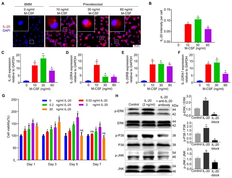
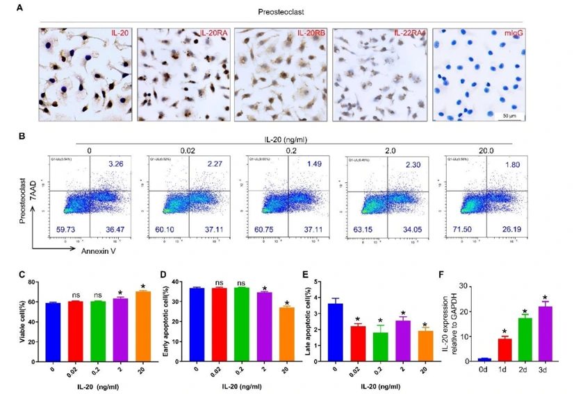
IL-20 Promoted Preosteoclast Proliferation by MAPK Pathways
Osteoimmunology mediators maintain bone homeostasis by coordinating bone formation and bone resorption processes. IL-20 is known to regulate expression of many osteoimmunology mediators; however, its role during bone remodeling has not been determined. Meng et al. determined IL-20 expression in remodeling alveolar bone during OTM and its association with osteoclast function.
To characterize IL-20 expression and involvement in preosteoclast differentiation, BMMs were treated to induce preosteoclasts which were then stained by immunofluorescence and immunohistochemistry. BMMs treated with M-CSF had increased expression of IL-20 in preosteoclasts as determined by immunofluorescence intensity. Maximum fluorescence intensity occurred when cells were treated with 30 ng/mL M-CSF (Fig. 1A, B). qRT-PCR results demonstrated M-CSF alters expression of IL-20 and IL-20 receptors (IL-20RA, IL-20RB, IL-22RA1) in preosteoclasts (Fig. 1C-F). Time-dependent increases in IL-20 expression were observed when treated with 30 ng/mL M-CSF (Fig. 2F), which was confirmed by immunohistochemistry (Fig. 2A). CCK8 assays and flow cytometry analyses showed IL-20 promotes proliferation of preosteoclasts in a dose-dependent manner and inhibits apoptosis (Fig. 1G and Fig. 2B-E). Western blot results indicate IL-20 induces MAPK signaling (ERK, p38, JNK) which was partially inhibited by anti-IL-20 antibodies (Fig. 1H). In conclusion, IL-20 is expressed in M-CSF-induced preosteoclasts and potentiates their function through MAPK signaling.
Ask a Question
Write your own review

Description: The thoracic aorta is located in the chest cavity and gives off arteries that branch to the esophagus, pericardium, lungs, and trachea. The thoracic aorta can be subdivided into the ascending aorta, ...

Description: Rat Podocytes are isolated from normal rat kidney. The cells are characterized by immunofluorescence with antibodies specific to podocin, Ang1, Nephrin, ACTN4, NPHS2. T25 flasks is required for cell ...

Description: Rat Bronchial Smooth Muscle Cells are isolated from normal rat bronchi tissue. Rat Bronchial Smooth Muscle Cells are characterized by immunofluorescence with antibodies specific to alpha-actin. T25 ...

Description: Guinea Pig Endothelial Cells from Creative Bioarray are isolated from guinea pig tissue. Prior to shipping, cells at passage 2 are detached from flasks and immediately cryopreserved in vials. Each ...

Description: Rat Lung Epithelial Cells are isolated from normal rat lung tissue. The cells are characterized by immunofluorescence with antibodies specific to CK-18, CK-19. T25 flasks is required for cell ...

Description: Rat Vein Endothelial Cells from Creative Bioarray are isolated from inferior vena cava tissue of 6-8 week old laboratory Sprague-Dawley rat. Rat Vein Endothelial Cells are grown in T75 tissue culture ...