
Human Tonsil Fibroblasts (HTF)
Cat.No.: CSC-7705W
Species: Human
Source: Tonsil
Cell Type: Fibroblast
- Specification
- Background
- Scientific Data
- Q & A
- Customer Review
Human Tonsil Fibroblasts (HTF) are primary stromal fibroblasts obtained from human palatine tonsil tissue, which is a secondary lymphoid organ that plays a role in mucosal immunity and immunological surveillance. HTF are an important part of the tonsillar microenvironment, contributing to the structural organization and functional maintenance of lymphoid tissues. They produce extracellular matrix proteins, cytokines, chemokines and growth factors that govern immune cell trafficking and activation.
Human Tonsil Fibroblasts are spindle-shaped cells characteristic of stromal cells and express markers associated with fibroblastic reticular cells (FRCs) and lymphoid tissue fibroblasts. These cells actively interact with T lymphocytes, B lymphocytes, dendritic cells and other immune populations, helping to establish specialized immune niches that facilitate antigen presentation, germinal center formation and adaptive immune responses. Recent investigations have shown that tonsillar stromal fibroblasts play an important role in controlling immune cell location, tissue homeostasis and inflammatory responses in the upper respiratory tract.
HTF are an invaluable in vitro model for studying lymphoid stromal biology, immune cell-stromal interactions, chronic inflammation, infectious illnesses and mucosal immunological processes. They are increasingly used in research of lymphoid organ development, vaccination responses, immunotherapy and modified lymphoid tissue models. These Fibroblasts provide a physiologically appropriate platform to advance studies in immunology, regenerative medicine and translational treatments since they mimic important aspects of the human tonsillar milieu.
Inflammation Reshapes Human PI16⁺ Fibroblastic Reticular Cells to Promote Immune–Stromal Crosstalk
Fibroblastic reticular cells (FRCs) organize lymphoid tissue microenvironments, yet their age‑ and inflammation‑related changes in humans are unclear. Martine et al. profiled tonsillar stromal cells across the lifespan, focusing on peptidase inhibitor 16–expressing reticular cells (PI16⁺ RCs).
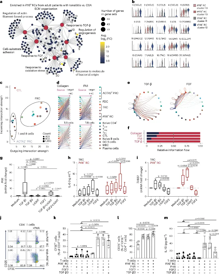
PI16⁺ RCs showed minimal age‑associated transcriptional change but pronounced activation during inflammation (tonsillitis vs. obstructive sleep apnea controls), including upregulation of angiogenic and niche‑forming gene sets such as FN1and response‑to‑bacterial‑origin pathways (Fig. 1a, b). Single‑cell RNA‑seq of matched T/B cells combined with Cell Chat analysis identified FRCs as highly interactive stromal components, exhibiting strong autocrine signaling and predicted forward signaling to lymphocytes via collagen, fibronectin (FN1), CXCL, THBS, IL‑6, and ICAM1 (Fig. 1c–e). Inflammation substantially increased intercellular communication network density across FRC subsets (Fig. 1f).
In vitro validation confirmed that TGF‑β1 downregulated PI16 and upregulated FN1in tonsillar fibroblasts, mimicking the inflamed PI16⁺ RC profile (Fig. 1g). TGF‑β1 and LIGHT synergistically or additively induced IL‑6 secretion and ICAM1 surface expression, whereas FGF2 antagonized these pro‑inflammatory effects.
These findings demonstrate that human tonsillar PI16⁺ RCs are dynamically remodeled by inflammation, integrating reciprocal T/B cell signals (TGF‑β, LIGHT) and FGF modulation to regulate stromal phenotype, cytokine output, and immune cell recruitment within secondary lymphoid tissue.
Ask a Question
Write your own review
Description: HTonEpiC from Creative Bioarray are isolated from human normal tonsil tissue. HTonEpiC are cryopreserved at passage one culture and delivered frozen. Each vial contains >5 x 10^5 cells in 1 ml ...
Description: Primary Human Tonsil Microvascular Endothelial Cells were initiated from normal human tonsil tissue by elutriation.These cells were originated using Complete Serum-Free Medium Kit With ...