

Immortalized Human Hepatic Stellate Cells-SV40T
Cat.No.: CSC-I2096Z
Species: homo sapiens
Morphology: Polygonal
Culture Properties: Adherent
- Specification
- Background
- Scientific Data
- Q & A
- Customer Review
- Documents
Note: Never can cells be kept at -20°C.
Immortalized Human Hepatic Stellate Cells (HSCs) engineered via stable expression of the SV40 Large T antigen (SV40T) represent a robust, genetically defined experimental platform that overcomes the finite lifespan, inter-donor variability, and ethical constraints inherent to primary human HSC cultures. By overriding cell-cycle arrest through SV40T-mediated inactivation of p53 and pRB pathways, these cells achieve unlimited proliferative capacity while retaining the activated myofibroblastic phenotype characteristic of pathological HSC transdifferentiation.
A principal advantage of SV40T-immortalized human HSCs is their faithful preservation of key functional and molecular hallmarks. These cells consistently express canonical activation markers including α-smooth muscle actin (α-SMA), vimentin, glial fibrillary acidic protein (GFAP), platelet-derived growth factor receptor-β (PDGFRβ), discoidin domain receptor 2 (DDR2), and the leptin receptor (Ob-R). Functionally, they secrete type I procollagen, pro-MMP-2, MT1-MMP, TIMP-1, and TIMP-2, and retain the capacity to absorb, accumulate, and esterify retinol—mimicking critical aspects of native HSC biology in hepatic fibrogenesis and retinoid metabolism.
These cells exhibit robust responsiveness to canonical fibrogenic stimuli: TGF-β1 potently upregulates collagen expression, while PDGF-B drives proliferation. Their high transfection efficiency further enables mechanistic studies involving gene overexpression, knockdown, and CRISPR/Cas9-mediated genome editing. Authenticated by STR profiling and extensively characterized in peer-reviewed literature, SV40T-immortalized human HSCs provide a standardized, reproducible, and scalable resource for investigating liver fibrosis pathogenesis, anti-fibrotic drug screening, extracellular matrix dynamics, and cytokine signaling pathways.
In Vitro Modeling of Liver Fibrosis with 3D Co-culture System Using a Novel Human Hepatic Stellate Cell Line
To develop an in vitro model that more reliably recapitulates the in vivo regenerative/supportive functions of hepatic stellate cells (HSCs) and TGF-beta pathway dependency on fibrosis progression, we aimed to establish a novel human HSC line and apply it to better mimic the in vivo 3D culture conditions.
In this study, Lee, Ho‐Joon generated an immortalized human HSC line, LSC-1, using SV-40 and hTERT, and characterized it using various HSC markers. they also compared the function of the LSC-1 line at the early regeneration stage, transdifferentiation level change, and TGF-β1 responsiveness in 3D hepatic co-culture formats with those of existing in vitro models, namely LX-2 cells. LSC-1 supported albumin production and hepatic differentiation state of co-cultured HepaRG in different 3D culture conditions, providing a robust starting point and timeframe for fibrosis modeling. LSC-1 also showed better responsiveness to a repeated dose of TGF-β1 in the 3D spheroid model. This new LSC-1 line may provide alternative options for in vitro mechanistic and clinical hepatic disease modeling studies.
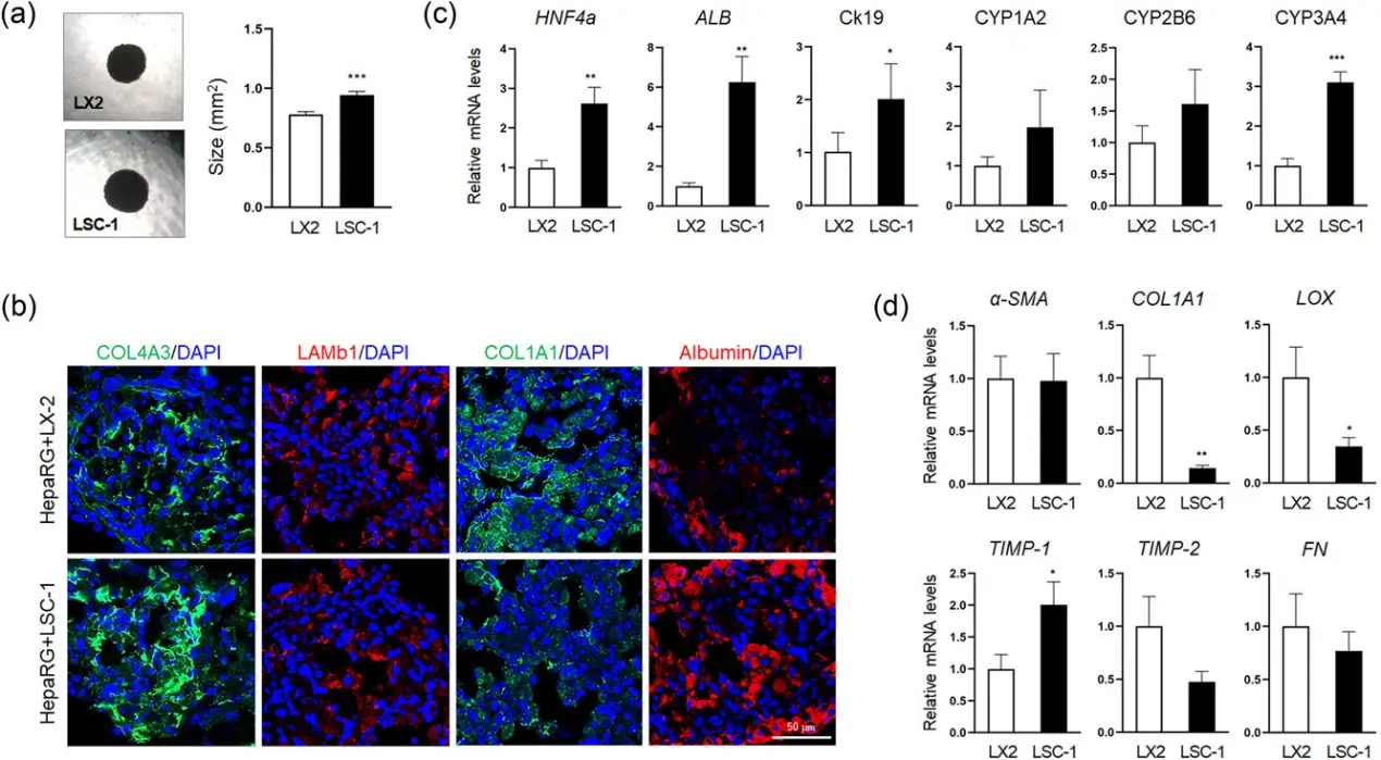
Hepatic stellate cells are critical for studying liver biology and pathophysiology. They are involved in processes such as liver fibrosis, inflammation, and regeneration. Research on these cells helps in understanding liver diseases, including cirrhosis and non-alcoholic fatty liver disease (NAFLD), and developing therapeutic strategies.
Immortalized Human Hepatic Stellate Cells can be utilized in various research applications, including:
Investigating the mechanisms of liver fibrosis and tissue remodeling.
Testing the effects of drugs and compounds on stellate cell activation.
Studying the interactions between stellate cells and other liver cell types (e.g., hepatocytes, Kupffer cells).
Exploring the role of hepatic stellate cells in liver inflammation and regeneration.
Ask a Question
Write your own review
 Immortalized Human Hepatic Stellate Cells-SV40T
Description: Nasal epithelial cells form the outermost protective layer against environmental factors. They clean, humidify, and warm inhaled air. They produces mucus, which binds particles that are subsequently ...
Description: Immortalized Human Splenic Endothelial Cells-SV40 have been obtained immortalizing Human Splenic Endothelial Cells with SV40LT expressing lentiviral particles. Immortalized cells were controlled ...

Description: Immortalized Human Corneal Epithelial Cells-SV40 have been obtained immortalizing Human Corneal Epithelial Cells with Lenti-SV40 Lentivirus. Immortalized cells were controlled passaging side by side ...
Description: Immortalized Human Lymphatic Endothelial Cells-SV40 were developed from human tissues transduced with a lentiviral expression vector containing the SV40T gene. The cell line was continuously cultured ...
Description: Immortalized Human Retinal Pigment Epithelial Cells were isolated from neonatal human globes and spontaneously immortalized in culture bypassing crisis. They retained typical morphology of the ...
- Adipose Tissue-Derived Stem Cells
- Human Neurons
- Mouse Probe
- Whole Chromosome Painting Probes
- Hepatic Cells
- Renal Cells
- In Vitro ADME Kits
- Tissue Microarray
- Tissue Blocks
- Tissue Sections
- FFPE Cell Pellet
- Probe
- Centromere Probes
- Telomere Probes
- Satellite Enumeration Probes
- Subtelomere Specific Probes
- Bacterial Probes
- ISH/FISH Probes
- Exosome Isolation Kit
- Human Adult Stem Cells
- Mouse Stem Cells
- iPSCs
- Mouse Embryonic Stem Cells
- iPSC Differentiation Kits
- Mesenchymal Stem Cells
- Immortalized Human Cells
- Immortalized Murine Cells
- Cell Immortalization Kit
- Adipose Cells
- Cardiac Cells
- Dermal Cells
- Epidermal Cells
- Peripheral Blood Mononuclear Cells
- Umbilical Cord Cells
- Monkey Primary Cells
- Mouse Primary Cells
- Breast Tumor Cells
- Colorectal Tumor Cells
- Esophageal Tumor Cells
- Lung Tumor Cells
- Leukemia/Lymphoma/Myeloma Cells
- Ovarian Tumor Cells
- Pancreatic Tumor Cells
- Mouse Tumor Cells