
VCAP
Cat.No.: CSC-C8204L
Species: Homo sapiens (Human)
Source: Bone Metastasis
Morphology: epithelial
Culture Properties: adherent
- Specification
- Background
- Scientific Data
- Q & A
- Customer Review
CSF1PO: 10,12
D13S317: 11,12
D16S539: 9
D5S818: 12
D7S820: 9,12
THO1: 9.3
TPOX: 8,11
vWA: 18,19
VCaP (Vertebral Cancer of the Prostate) cell line is a well-characterized in vitro model, derived from a vertebral metastasis of castration-resistant prostate cancer (CRPC). First established in 1997, this cell line has been extensively studied and possesses some notable characteristics that make it a valuable tool for prostate cancer research.
VCaP is an epithelial adherent cell line with moderate growth kinetics and a doubling time of 48-72 hours under standard culture conditions. The VCaP cell line shows overexpression of both wild-type androgen receptor (AR) and estrogen receptor alpha (ERα). This unique property makes VCaP an attractive model to study both androgen and estrogen signaling in prostate cancer. In addition to the AR and ERα expression, the VCaP cells also harbor the TMPRSS2-ERG gene fusion, which is present in approximately 50% of clinical prostate cancer cases.
Originating from a metastatic lesion in the bone, VCaP is particularly useful for studying the mechanisms underlying CRPC development and bone metastasis. The cells have been employed to study AR signaling dynamics, therapeutic resistance mechanisms, and tumor-microenvironment interactions. VCaP cells can also form tumors in xenograft models.
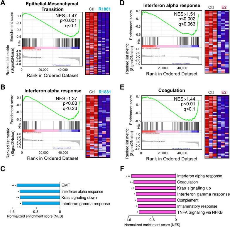
Bulk mRNA-Sequencing Data of the Estrogen and Androgen Responses in the Human Prostate Cancer Cell Line VCaP
Estrogen signaling in prostate cancer (PCa) is under-investigated compared to well-established androgen dependence. VCaP cells, expressing wild-type AR and ERα, are ideal for investigating these hormonal interactions. Lafront et al. generated a transcriptomic atlas of androgen and/or estrogen-dependent transcriptional changes in VCaP cells using bulk mRNA-seq.
VCaP cells were treated with androgens and/or estrogens for 24 h, mRNA purified, sequenced, and aligned to the human transcriptome. Transcriptomic changes were analyzed after quantification. The raw data were submitted to the Gene Expression Omnibus (GEO) repository and can be found using the GSE256370 reference number. The dataset contains 12 samples (exact number of four treatment groups: control, R1881 [synthetic androgen], estradiol and estradiol + R1881, with three replicates per group) and, here, were used to test the downregulated transcriptional response upon stimulation by androgen and estrogen with the GSEA tool (Fig. 1).
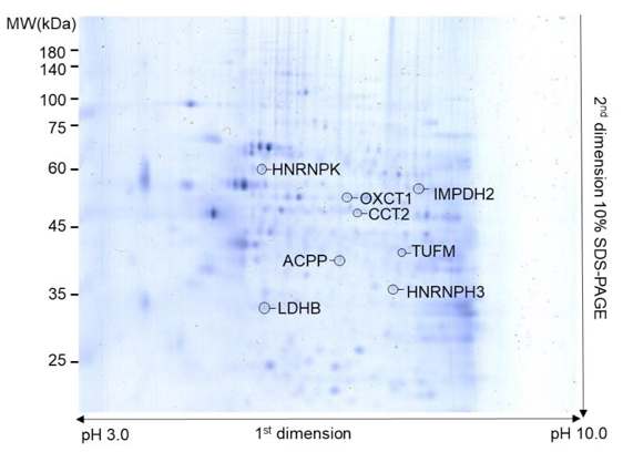
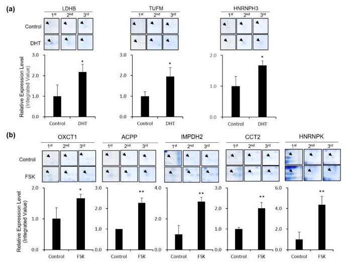
Androgen- and PKA Signaling-Induced Changes in the Proteomic Profile of VCaP Cells and Identification of Differentially Expressed Proteins
Androgen signaling is crucial in prostate cancer, but resistance leads to CRPC, where PKA may bypass androgen dependence. The key proteins involved remain unknown. You’s team aims to identify proteins regulated by androgen and PKA signaling in VCaP prostate cancer cells, uncovering potential molecular mechanisms behind CRPC progression and therapeutic targets.
VCaP cells were treated with androgen or forskolin (activator of PKA) then subjected to proteomic analysis to quantify the differentially expressed proteins. In total, 113 protein spots that have different expression between the control and DHT or FSK were detected. Among these, eight spots display significant changes in density (> 1.5-fold changes in expression, p < 0.05). These eight differentially expressed proteins (Fig. 2) were significantly increased in response to DHT or FSK in three repeat experiments (Fig. 3). The proteins were subsequently identified by MS analyses. Three proteins were found to be altered in VCaP cells in response to DHT: LDHB, TUFM and HNRNPH3. Five proteins were specifically increased in response to FSK: OXCT1, ACPP, IMPDH2, CCT2 and HNRNPK. To further understand the functional implication of these proteins, they performed a Gene Ontology (GO) analysis on the cellular location and biological function of the proteins. The GO analysis revealed that all the identified proteins are involved in the metabolic process. Notably, metabolic reprogramming has been linked to AR signaling re/activation and antagonism that drives the progression of CRPC.
Ask a Question
Write your own review
- You May Also Need
Description: C4-2 is a cell line with epithelial-like morphology that was isolated from a human prostate cancer LNCaP cell subcutaneous xenograft tumor of castrated mouse.
Description: Cytogenetic analysis and DNA fingerprinting at the Creative Bioarray documented that this cell line is identical with the human prostate carcinoma-derived cell line DU-145, but shows some ...

Description: 22Rv1 is a human prostate carcinoma epithelial cell line derived from a xenograft that was serially propagated in mice after castration-induced regression and relapse of the parental, ...

Description: prostate epithelial cells from a 68-year-old man with benign prostate hyperplasia; cells were immortalized with SV-40 large T-antigen; cells were described to express cytokeratins 8, 18, and 19 (but ...

Description: Derived from a human prostate carcinoma xenograft (CWR22R) that was serially propagated in nude mice after castration-induced regression and relapse of the parental, androgen-dependent CWR22 xenograft
- Adipose Tissue-Derived Stem Cells
- Human Neurons
- Mouse Probe
- Whole Chromosome Painting Probes
- Hepatic Cells
- Renal Cells
- In Vitro ADME Kits
- Tissue Microarray
- Tissue Blocks
- Tissue Sections
- FFPE Cell Pellet
- Probe
- Centromere Probes
- Telomere Probes
- Satellite Enumeration Probes
- Subtelomere Specific Probes
- Bacterial Probes
- ISH/FISH Probes
- Exosome Isolation Kit
- Human Adult Stem Cells
- Mouse Stem Cells
- iPSCs
- Mouse Embryonic Stem Cells
- iPSC Differentiation Kits
- Mesenchymal Stem Cells
- Immortalized Human Cells
- Immortalized Murine Cells
- Cell Immortalization Kit
- Adipose Cells
- Cardiac Cells
- Dermal Cells
- Epidermal Cells
- Peripheral Blood Mononuclear Cells
- Umbilical Cord Cells
- Monkey Primary Cells
- Mouse Primary Cells
- Breast Tumor Cells
- Colorectal Tumor Cells
- Esophageal Tumor Cells
- Lung Tumor Cells
- Leukemia/Lymphoma/Myeloma Cells
- Ovarian Tumor Cells
- Pancreatic Tumor Cells
- Mouse Tumor Cells