
KYSE-520
Cat.No.: CSC-C0419
Species: Homo sapiens (Human)
Source: Esophagus
Morphology: epitheloid cells growing adherently as monolayers, occasional giant cells present (ca. 1%)
Culture Properties: monolayer
- Specification
- Background
- Scientific Data
- Q & A
- Customer Review
Immunology: cytokeratin +, cytokeratin-7 -, cytokeratin-8 +, cytokeratin-17 +, cytokeratin-18 +, cytokeratin-19 +, desmin -, endothel -, EpCAM +, GFAP -, neurofilament -
KYSE-520 is a well-established human esophageal squamous cell carcinoma (ESCC) cell line, originally derived from a primary tumor of a 66-year-old male patient in the 1990s. It retains key molecular and phenotypic features of ESCC, including epithelial morphology, proliferation as an adherent monolayer, and characteristic genetic alterations such as TP53 mutations and CDKN2A deletions, which are common drivers of esophageal cancer pathogenesis. These cells also exhibit dysregulated signaling pathways (e.g., EGFR, PI3K/AKT) and express markers like cytokeratins, making them valuable for studying mechanisms of tumorigenesis, metastasis, and drug response.
KYSE-520 cells are moderately differentiated and useful for studying epithelial-mesenchymal plasticity (EMP) in esophageal cancer. They exhibit heterogeneity, consisting of both epithelial-like (CD44v+) and mesenchymal-like (CD44v-) subpopulations. These two populations are able to interconvert, reflecting a dynamic EMP process. This property makes KYSE520 a good model for studying ESCC cancer stem cell properties and mechanisms of chemoresistance.
KYSE520 cells have been used to explore the role of fibroblast growth factor receptor-like 1 (FGFRL1) in therapeutic studies. Studies have shown that FGFRL1-deficient KYSE520 cells exhibit reduced tumor growth and motility, as well as a decrease in matrix metalloproteinase-1 (MMP-1) and fibroblast growth factor binding protein 1 (FGFBP1) expression. These findings highlight the importance of FGFRL1 in tumorigenesis and suggest potential therapeutic targets. In addition, the EMP dynamics and related molecular pathways in KYSE520 cells provide insights into the mechanisms of ESCC progression and resistance, contributing to the development of targeted therapies.
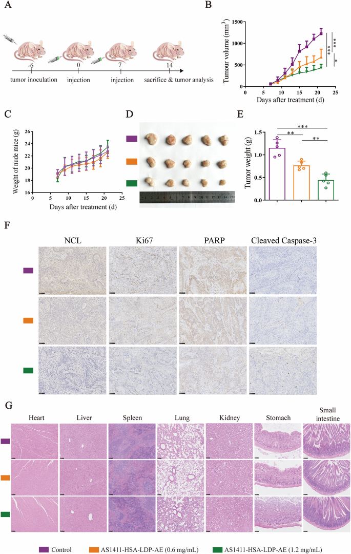
A new NCL-targeting aptamer-drug conjugate as a promising therapy against esophageal cancer
Background
Esophageal cancer (EC) is a highly lethal tumor of the digestive system. Nucleolin (NCL) is overexpressed in various tumors and is a potential therapeutic target. In this study, a novel NCL-targeting aptamer-drug conjugate AS1411-HSA-LDP-AE was developed. It consists of the AS1412 aptamer, human serum albumin, leucidomycin de-acylated protein, and an active enediynone chromophore. The results showed that this conjugate could specifically bind and internalize into esophageal cancer cells. It targeted and accumulated in tumors in the xenograft model and exhibited strong cytotoxic activity, inhibition of migration and invasion, induction of apoptosis, and significant in vivo anti-tumor effects. This provides a promising strategy for targeted therapy of esophageal cancer.
Key Findings
This study successfully constructed an aptamer-drug conjugate AS1411-HSA-LDP-AE targeting nucleolin (NCL). NCL is overexpressed in esophageal cancer cells, with the highest expression in KYSE520. AS1411-HSA-LDP can bind to esophageal cancer cells in a concentration-dependent manner and is efficiently internalized. In KYSE520, it mainly undergoes macropinocytosis. In the xenograft model, this conjugate can long-term target and accumulate at the tumor site. AS1411-HSA-LDP-AE strongly inhibits the proliferation of esophageal cancer cells, with the IC50 being the lowest in KYSE520, and it inhibits migration and invasion and induces apoptosis. The apoptotic mechanism involves the mitochondrial pathway: increased ROS, decreased mitochondrial membrane potential, release of cytochrome c, upregulation of Bax, downregulation of Bcl-2, and inhibition of PARP1. In vivo experiments showed that AS1411-HSA-LDP-AE significantly inhibited tumor growth. In the KYSE520 model, the inhibition rates at 0.6 and 1.2 mg/kg doses were 49.9% and 70.1%, respectively, and at 1.6 mg/kg dose in the EC109 model, the inhibition rate was 53.8%. There was no significant toxicity. Immunohistochemistry showed that NCL and Ki-67 were downregulated, and cleaved caspase-3 was upregulated. These results indicate that AS1412-HSA-LDP-AE is a promising targeted therapeutic drug for esophageal cancer.
Contribution
Creative Bioarray offers human esophageal cancer cell lines KYSE520 and KYSE70, which are used for in vitro experiments and the establishment of in vivo xenograft models in this study. The availability of these cell lines is crucial for evaluating the targeting, uptake mechanism and anti-tumor effect of AS1411-HSA-LDP-AE, providing essential material support for the success of the experiments in this study.
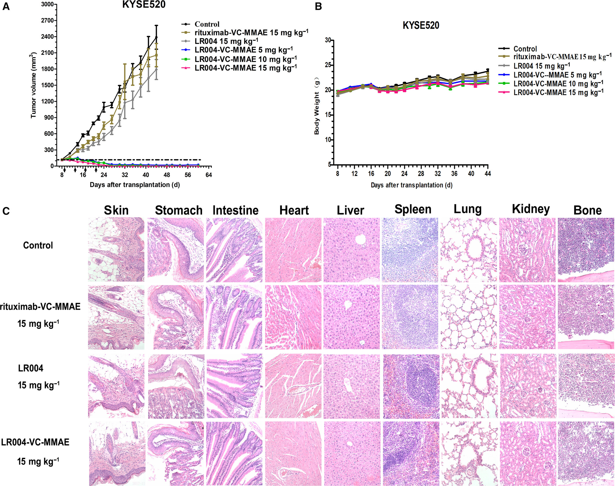
An EGFR-targeting antibody–drug conjugate LR004-VC-MMAE: potential in esophageal squamous cell carcinoma and other malignancies
Background
This study developed LR004-VC-MMAE, an anti-EGFR antibody–drug conjugate linking LR004 to monomethyl auristatin E via a VC linker. LR004 showed high EGFR affinity, internalization, and lysosomal trafficking. The ADC had approximately 4.0 drug–antibody ratio, retained EGFR binding, and killed EGFR-positive cancer cells at nanomolar concentrations. In KYSE520 esophageal squamous cell carcinoma and A431 xenografts, LR004-VC-MMAE caused marked tumor regression, including complete regression at 15 mg/kg, whereas naked LR004 had little effect. It also showed favorable pharmacokinetics and safety, supporting further development for ESCC and other EGFR-expressing malignancies.
Key Findings
LR004-VC-MMAE was prepared by conjugating MMAE to LR004 through a VC linker, with an average drug–antibody ratio of ~4.0. It retained EGFR binding comparable to LR004 (KD 1.313 nM vs 0.916 nM), was rapidly internalized, and colocalized with lysosomal LAMP-1. In vitro, LR004-VC-MMAE was highly cytotoxic to EGFR-high ESCC, NSCLC, breast, pancreatic, and A431 cells, with IC50 values in the nanomolar range, while EGFR-negative Karpas 299 cells and rituximab-VC-MMAE controls were insensitive. It induced apoptosis and G2/M arrest in ESCC and A431 cells. In vivo, LR004-VC-MMAE significantly inhibited KYSE520 and A431 xenografts. At 5 mg/kg it showed significant activity, and at 15 mg/kg it achieved complete tumor regression in KYSE520 after four injections, with no recurrence until day 60. It also inhibited large A431 tumors (400–500 mm3). Naked LR004 had minimal antitumor effect. Pharmacokinetic analysis showed a total antibody half-life of ~113.6 h and low free MMAE, indicating ADC stability and a favorable safety profile.
Contribution
Creative Bioarray provided the human esophageal squamous cell carcinoma KYSE520 cell line used in this study. This EGFR-overexpressing line enabled in vitro cytotoxicity, binding, internalization, and lysosomal trafficking assays, and supported the in vivo KYSE520 xenograft efficacy study, helping demonstrate the potent antitumor activity of LR004-VC-MMAE.
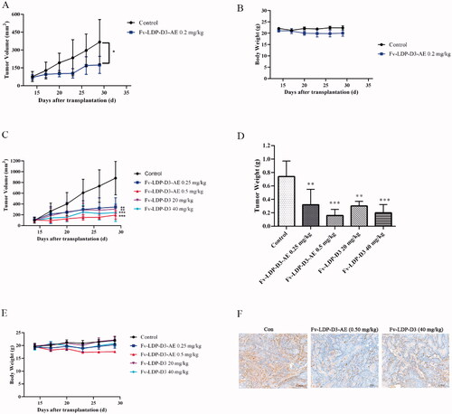
A recombinant scFv antibody-based fusion protein that targets EGFR associated with IMPDH2 downregulation and its drug conjugate show therapeutic efficacy against esophageal cancer
Background
This study evaluated EGFR-targeted recombinant fusion protein Fv-LDP-D3 and its antibody-drug conjugate Fv-LDP-D3-AE against esophageal cancer. Fv-LDP-D3, composed of anti-EGFR Fv, lidamycin apoprotein LDP, and human serum albumin domain III, bound EGFR-overexpressing esophageal cancer cells, inhibited EGFR phosphorylation, downregulated IMPDH2, and entered cells via macropinocytosis. Fv-LDP-D3-AE integrated lidamycin enediyne chromophore, causing potent cytotoxicity, DNA damage, G2/M arrest, apoptosis, mitochondrial damage, and inhibiting migration/invasion. Both agents accumulated in tumors and suppressed xenograft growth at tolerated doses, suggesting promising targeted therapy candidates.
Key Findings
Fv-LDP-D3 bound EGFR-overexpressing KYSE520/KYSE150 esophageal cancer cells in a concentration-dependent manner and showed weak binding to NIH3T3. It was internalized through macropinocytosis and inhibited proliferation, EGFR phosphorylation, and IMPDH2 expression. Fv-LDP-D3 induced apoptosis and accumulated specifically in tumor sites with retention up to 26 days. Fv-LDP-D3-AE displayed potent cytotoxicity across esophageal cancer cell lines, suppressed migration/invasion, induced DNA damage (p-Histone H2A.X up, Chk1 down), G2/M arrest, apoptosis, and mitochondrial damage. In KYSE150 xenografts, Fv-LDP-D3-AE at 0.2 mg/kg achieved 64% tumor inhibition. In KYSE520 xenografts, Fv-LDP-D3 at 20/40 mg/kg yielded 73%/81% inhibition, while Fv-LDP-D3-AE at 0.25/0.5 mg/kg yielded 69%/89% inhibition, with no significant toxicity or histopathological changes. Ki-67 was reduced. These results support EGFR-targeted Fv-LDP-D3 and its ADC as promising esophageal cancer therapeutics.
Contribution
Creative Bioarray supplied KYSE150 and KYSE520 human esophageal squamous cell carcinoma cell lines. These cells were essential for in vitro binding, cytotoxicity, and mechanism studies and for establishing KYSE520/KYSE150 xenograft models used to evaluate Fv-LDP-D3 targeting, IMPDH2 downregulation, and Fv-LDP-D3-AE antitumor efficacy. No other contributions were disclosed.
TK-642 Inhibits Cell Proliferation and Induces Apoptosis of Esophageal Cancer Cells In Vitro
Src homology-2-containing protein tyrosine phosphatase 2 (SHP2) is a promising therapeutic target for cancer therapy. Pyrazolopyrazine-based TK-642 is identified as a highly potent, selective, orally bioavailable allosteric SHP2 inhibitor with favorable pharmacokinetic profiles.
The cellular thermal shift assay (CETSA) was performed to investigate the potential of TK-642 in targeting intracellular SHP2 protein. As illustrated in Fig. 1A, a significantly higher expression level of SHP2 protein was observed within the temperature range of 45-63°C in EGFR amplified KYSE-520 esophageal cancer cell lines treated with TK-642, as compared to the DMSO treatment group. This finding indicated that TK-642 could effectively bind to and enhance the thermal stability of intracellular SHP2 protein in KYSE-520 cells.
Considering the well-known oncogenic roles of SHP2 in the development of esophageal cancer, we proceeded to investigate the impact of TK-642 on cell proliferation and apoptosis of KYSE-520 cell lines, with SHP099 serving as a control. The results revealed that both compounds exhibited moderate effects on the proliferation of KYSE-520 cells. Especially, the inhibitory activity of TK-642 on the KYSE-520 cells was approximately 3-fold greater than that of SHP099 (Fig. 1B). Moreover, we also verified the anti-tumor activity of TK-642 in non-small cell lung cancer H1975 and gastric cancer HGC-27 cells (Fig. 1C). Similar to SHP099, TK-642 also induced the apoptosis of KYSE-520 cells in a concentration-dependent manner (Fig. 1D).
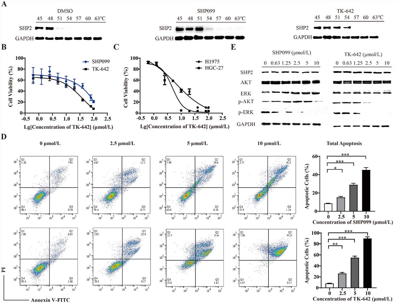 Fig. 4. TK-642 inhibits cell proliferation and induces apoptosis of esophageal cancer cells KYSE-520 (Tang, Kai, et al. 2024).
Cell Proliferation Was Reduced in Hsa_circ_0001615 Knockdown Cells
Recent studies have found that circular RNAs (circRNAs) play important roles in tumorigenesis. This study aimed to determine the function and potential mechanisms of hsa_circ_0001615 in esophageal cancer.
The endogenous expression of hsa_circ_0001615 was quantified in three esophageal cancer cell lines (ECA-109, TE-1, KYSE520) and normal esophageal cells (HEEC). The relative expression of hsa_circ_0001615 was higher in the three esophageal cancer cell lines than in normal esophageal cells. Moreover, it was higher in ECA-109 and KYSE520 cells than in TE-1 cells (Fig. 2A).
ECA-109 and KYSE520 cell lines were used to transfect hsa_circ_0001615 siRNA to gain a better understanding of the functions of hsa_circ_0001615. The relative expression of hsa_circ_0001615 was significantly reduced after transfection with siRNAs for 48 h (Fig. 2B). Cell proliferation was significantly impaired when hsa_circ_0001615 was knocked down in ECA-109 and KYSE520 cells, as demonstrated by MTS assays (Fig. 2C). Cloning assay was also performed to confirm the growth defect in hsa_circ_0001615 knockdown cell lines. The number of clone formation was substantially decreased when hsa_circ_0001615 was knocked down (Fig. 2D). These results indicated that reduced hsa_circ_0001615 expression decreased cell proliferation and colony formation in ECA-109 and KYSE520 cell lines.
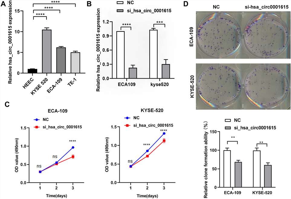 Fig. 5. Reduced cell proliferation in hsa_circ_0001615 knockdown cells (Dai, Yukai, et al. 2024).
Ask a Question
Write your own review
- You May Also Need
Description: Human moderately differentiated squamous cell carcinoma cell line established from esophageal cancer.
Description: Human moderately differentiated squamous cell carcinoma cell line established from esophageal cancer.
Description: Human moderately differentiated squamous cell carcinoma cell line established from esophageal cancer.
Description: Human squamous cell carcinoma from oral cavity via mouse transplantation.
Description: Human squamous cell carcinoma cell line established from recurrent cancer (esophageal cancer).
- Adipose Tissue-Derived Stem Cells
- Human Neurons
- Mouse Probe
- Whole Chromosome Painting Probes
- Hepatic Cells
- Renal Cells
- In Vitro ADME Kits
- Tissue Microarray
- Tissue Blocks
- Tissue Sections
- FFPE Cell Pellet
- Probe
- Centromere Probes
- Telomere Probes
- Satellite Enumeration Probes
- Subtelomere Specific Probes
- Bacterial Probes
- ISH/FISH Probes
- Exosome Isolation Kit
- Human Adult Stem Cells
- Mouse Stem Cells
- iPSCs
- Mouse Embryonic Stem Cells
- iPSC Differentiation Kits
- Mesenchymal Stem Cells
- Immortalized Human Cells
- Immortalized Murine Cells
- Cell Immortalization Kit
- Adipose Cells
- Cardiac Cells
- Dermal Cells
- Epidermal Cells
- Peripheral Blood Mononuclear Cells
- Umbilical Cord Cells
- Monkey Primary Cells
- Mouse Primary Cells
- Breast Tumor Cells
- Colorectal Tumor Cells
- Esophageal Tumor Cells
- Lung Tumor Cells
- Leukemia/Lymphoma/Myeloma Cells
- Ovarian Tumor Cells
- Pancreatic Tumor Cells
- Mouse Tumor Cells