
CGTH-W-1
Cat.No.: CSC-C0412
Species: Homo sapiens (Human)
Source: Thyroid Gland
Morphology: adherent large epitheloid cells with long processes growing in monolayers with multilayer foci
Culture Properties: monolayer
- Specification
- Background
- Scientific Data
- Q & A
- Customer Review
Immunology: cytokeratin -, cytokeratin-7 -, cytokeratin-8 -, cytokeratin-17 -, cytokeratin-18 -, des
CGTH-W-1 (formerly known as W-1) is a human thyroid carcinoma cell line first described as being established from the sternal metastasis of follicular thyroid carcinoma from an elderly woman. It is commonly used as an in vitro model of thyroid cancer, specifically follicular thyroid carcinoma and poorly differentiated thyroid carcinoma. Cells maintain adherent epithelial morphology and grow as monolayers. Cells have relatively fast doubling times of ~18-25 hours. Molecular analyses have identified driver mutations such as TP53 and TERT promoter mutations that are commonly seen in aggressive thyroid cancers. Initial characterization of the CGTH-W-1 cell line showed that they express insulin-like growth factor-1 receptors and utilize IGF-1 signaling pathways. This made CGTH-W-1 useful to study growth factor-mediated tumor progression in vitro.
CGTH-W-1 was recently used in studies along with FTC-133 and BcPAP thyroid cancer cell lines to study gene regulation and extracellular vesicle-mediated signaling as well as interactions with tumor microenvironments. They have also been used for proteomic analysis and functional analysis including spheroid formation and cytoskeletal dynamics under varying physical conditions. CGTH-W-1 has been reported to be misidentified or cross-contaminated cell line and is a derivative of SW579.
PROX1 Knock-Down CGTH-W-1 Cells and Its Effect on Cell Motility and Invasive Potential
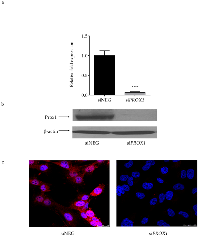
Prospero homeobox 1 (PROX1), a master regulator of lymphangiogenesis, is an implicated carcinogenic transcription factor. Its role in tumorigenesis and metastasis, however, is not well understood. In this study, Rudzińska et al. analyzed the role of PROX1 in thyroid tumorigenesis utilizing PROX1-siRNA-transfected follicular-derived thyroid cancer cell line CGTH-W-1. Effects of PROX1 depletion were analyzed on proliferation, cell cycle, apoptosis, and cell motility.
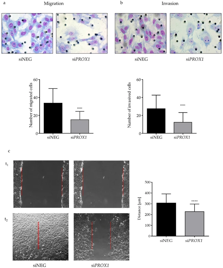
siRNA knockdown of PROX1 was validated by RT-qPCR, western blot, and immunocytochemistry. PROX1 transcript and protein levels were reduced by greater than 90% relative to non-silencing control siRNA-treated cells (Figure 1). Silencing of PROX1 led to statistically significant decreases in migration and invasion (>2-fold reduction, p < 0.0001) in consistently replicated experiments (Figure 2a, b).
Actinomycin-D-treated wound healing assays revealed PROX1 silencing significantly inhibited migration of cells (p = 0.0001), resulting in a 6.6-fold decrease in migrating cells with or without inhibition of proliferation (Figure 2c). Loss of PROX1 resulted in dramatic cytoskeletal reorganization visualized by immunofluorescence including alteration of cellular morphology and decreases in stress fibers and cell protrusions.
Ask a Question
Write your own review
- You May Also Need
Description: established from the thyroid gland from a 66-year-old male patient with thyroid gland anaplastic carcinoma

Description: Established in 1996 from the tumor tissue of a 57-year-old Caucasian man with local recurrence of a previously iodine-irradiated follicular thyroid cancer (well-differentiated with dispersed poorly ...

Description: established from the lymph node metastasis of a 63-year-old woman with anaplastic papillary thyroid carinoma; cells were described to not produce hormones, but to be partly positive for ...

Description: Established from cancer cells disseminated in the pleural fluid of a 44-year-old woman with undifferentiated giant cell carcinoma of the thyroid after chemotherapy, x-ray hyperthermia and OK432 ...
Description: Derived from a primary papillary thyroid carcinoma. Retains thyroid follicular cell differentiation e.g. thyroglobulin synthesis
- Adipose Tissue-Derived Stem Cells
- Human Neurons
- Mouse Probe
- Whole Chromosome Painting Probes
- Hepatic Cells
- Renal Cells
- In Vitro ADME Kits
- Tissue Microarray
- Tissue Blocks
- Tissue Sections
- FFPE Cell Pellet
- Probe
- Centromere Probes
- Telomere Probes
- Satellite Enumeration Probes
- Subtelomere Specific Probes
- Bacterial Probes
- ISH/FISH Probes
- Exosome Isolation Kit
- Human Adult Stem Cells
- Mouse Stem Cells
- iPSCs
- Mouse Embryonic Stem Cells
- iPSC Differentiation Kits
- Mesenchymal Stem Cells
- Immortalized Human Cells
- Immortalized Murine Cells
- Cell Immortalization Kit
- Adipose Cells
- Cardiac Cells
- Dermal Cells
- Epidermal Cells
- Peripheral Blood Mononuclear Cells
- Umbilical Cord Cells
- Monkey Primary Cells
- Mouse Primary Cells
- Breast Tumor Cells
- Colorectal Tumor Cells
- Esophageal Tumor Cells
- Lung Tumor Cells
- Leukemia/Lymphoma/Myeloma Cells
- Ovarian Tumor Cells
- Pancreatic Tumor Cells
- Mouse Tumor Cells