SNB-19
Cat.No.: CSC-C0391
Species: Homo sapiens (Human)
Source: Brain
Morphology: adherent fibroblastic cells growing as monolayer with contact inhibition, occasional giant cells
Culture Properties: monolayer
- Specification
- Background
- Scientific Data
- Q & A
- Customer Review
Immunology: cytokeratin -, cytokeratin-7 -, cytokeratin-8 -, cytokeratin-17 -, cytokeratin-18 -, desmin -, endothel -, GFAP +, neurofilament -, vimentin +
Viruses: ELISA:
SNB-19 cell line – A glioma cell line isolated from a meningioma. They exhibit an epithelial or fibroblast-like morphology and adhere strongly to culture dishes or flasks in monolayers. Not only does this cell line have wild-type p53 protein, it also has MGMT (O6-methylguanine-DNA methyltransferase), glial marker protein GFAP (glial fibrillary acidic protein), stem cell marker CD133 and high EGFR (epidermal growth factor receptor). In addition, SNB-19 cells also lack the CDKN2A/p16 gene and mutate the PTEN gene, but retain the wild-type IDH1 (isocitrate dehydrogenase 1) gene.
SNB-19 cells possess invasive and migratory capabilities, capable of forming neurospheres in vitro exhibiting stem cell-like properties. These features make them invaluable in studying glioma invasion, migration mechanisms, and stem cell characteristics. Additionally, SNB-19 cells secrete plasminogen activator, an enzyme crucial in the invasion and migration processes of tumor cells. In nude mouse experiments, SNB-19 cells can form tumors, validating their tumorigenic properties. In scientific research, the SNB-19 cell line is widely used to investigate the biological characteristics of gliomas, including tumor invasiveness, migration, and responses to chemotherapeutic agents.
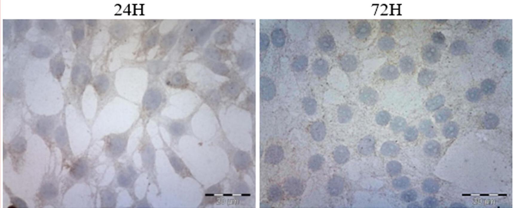 Fig. 1. Microscopic
images of SNB-19 after 24h and 72h of culture (Cierluk, K., Szlasa, W., et al., 2020).
Cepharanthine Induces ROS Stress in Glioma and Neuronal Cells Via Modulation of VDAC Permeability
Cepharanthine (CEP) is a bisbenzylisoquinoline alkaloid that may influence the occurrence and development of glioblastoma multiforme (GBM) through Voltage-dependent anion channels (VDAC). Cierluk's team investigated the biological effects of CEP on the glioblastoma cell line (SNB-19) and neuron cell line (PC-12 + NGF), finding that CEP induces ROS stress in glioma and neuronal cells by modulating VDAC permeability. Immunocytochemical staining showed that T-type calcium channel expression in SNB-19 cells increased with higher CEP concentrations, peaking at 10 μM. CEP did not affect T-type channel expression in PC-12 + NGF cells. For VDAC, SNB-19 cells showed a steady increase in expression with higher CEP concentrations, but no time-dependent changes. PC-12 + NGF cells had higher VDAC expression levels at higher CEP concentrations compared to SNB-19 cells, and their initial VDAC expression was significantly higher (Fig. 1). Cells incubated with increasing CEP concentrations showed a rise in ROS levels, especially in SNB-19 cells (Fig. 2A). Over time, even lower CEP concentrations led to greater ROS generation. Upon CEP addition, mitochondrial content dispersed evenly in the cytoplasm compared to the concentrated pattern in controls. In PC-12+NGF cells, ROS release was significant after 24h incubation at lower CEP concentrations, showing a distribution pattern similar to SNB-19 cells (Fig. 2B).
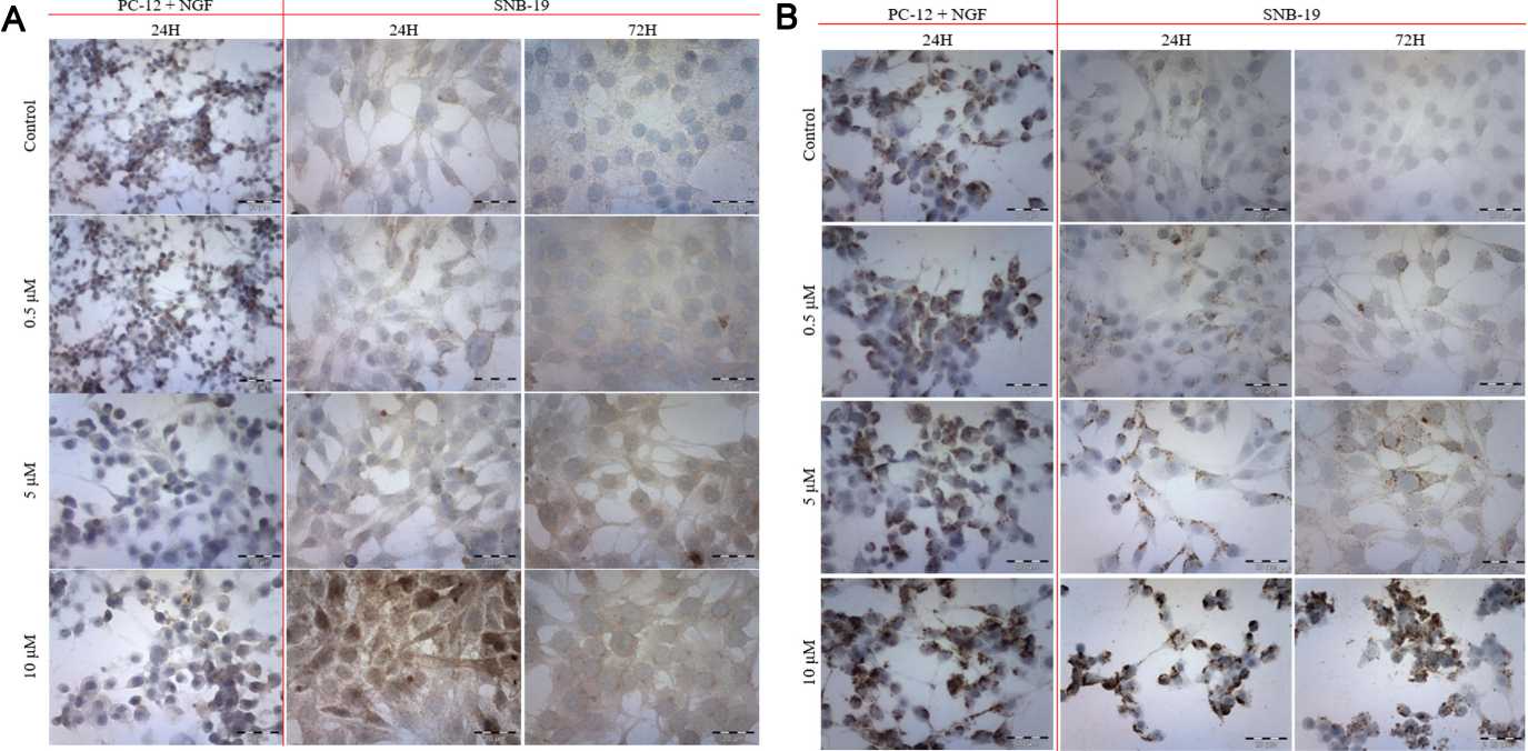 Fig.
1. The immunocytochemical evaluation of calcium T-type channel (A) and VDAC (B) with ABC technique after 24 h and 72
h incubation with varying concentrations of CEP (Cierluk, K., Szlasa, W., et al., 2020).
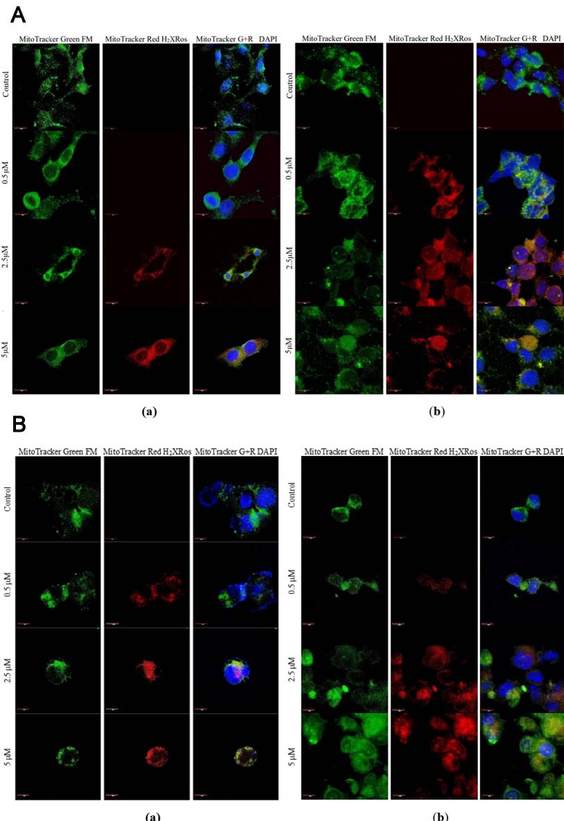 Fig.
2. Immunofluorescent staining of ROS in SNB-19 cells (A) and PC-12 + NGF cells (B) after (a) 24 h and (b) 72 h of
incubation with varying CEP concentrations (Cierluk, K., Szlasa, W., et al., 2020).
ZIKV Infection Reduced Mitochondrial Transmembrane Potential
Zika virus (ZIKV) causes neurodevelopmental disruption in newborn babies. Yang's team had studied how mitochondrial shape and function were altered in human NSCs and the human glioblastoma cell line SNB-19 following ZIKV infection. They explained how ZIKV infection disturbs mitochondrial dynamics, network organisation and activity by depleting MFN2 protein. In order to examine mitochondrial fragmentation under ZIKV infection, they speculated that dysregulation could be caused by a mismatch between mitochondrial fusion and fission proteins. In ZIKV-infected NSCs and SNB-19 cells, MFN2 protein levels fell after 24 hours even though infection was only 50 % (Fig. 3A-C). MFN1 reduction occurred in SNB-19 cells but not in NSCs, possibly due to lower neuronal expression (Fig. 3B). Immunofluorescence confirmed weaker MFN2 signals in infected cells compared to uninfected ones (Fig. 3D and E). Fragmented mitochondria were observed in ZIKV ENV-positive cells, indicating reduced mitochondrial fusion (Fig. 3F-H). Protein levels of OPA1, DNM1L, and FIS1 were unchanged, but DNM1L phosphorylation at Ser616, which promotes fission, was absent (Fig. 3B). This dephosphorylation could be a response to excessive fragmentation to curb further fission. Overall, ZIKV infection disrupted the balance of mitochondrial fusion and fission, leading to network fragmentation.
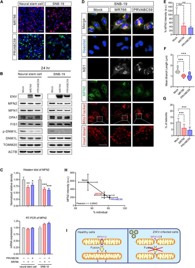 Fig. 3. MFN2 protein level was reduced in
ZIKV-infected cells (Yang S, Gorshkov K, et al., 2020).
Ask a Question
Write your own review
- You May Also Need
- Adipose Tissue-Derived Stem Cells
- Human Neurons
- Mouse Probe
- Whole Chromosome Painting Probes
- Hepatic Cells
- Renal Cells
- In Vitro ADME Kits
- Tissue Microarray
- Tissue Blocks
- Tissue Sections
- FFPE Cell Pellet
- Probe
- Centromere Probes
- Telomere Probes
- Satellite Enumeration Probes
- Subtelomere Specific Probes
- Bacterial Probes
- ISH/FISH Probes
- Exosome Isolation Kit
- Human Adult Stem Cells
- Mouse Stem Cells
- iPSCs
- Mouse Embryonic Stem Cells
- iPSC Differentiation Kits
- Mesenchymal Stem Cells
- Immortalized Human Cells
- Immortalized Murine Cells
- Cell Immortalization Kit
- Adipose Cells
- Cardiac Cells
- Dermal Cells
- Epidermal Cells
- Peripheral Blood Mononuclear Cells
- Umbilical Cord Cells
- Monkey Primary Cells
- Mouse Primary Cells
- Breast Tumor Cells
- Colorectal Tumor Cells
- Esophageal Tumor Cells
- Lung Tumor Cells
- Leukemia/Lymphoma/Myeloma Cells
- Ovarian Tumor Cells
- Pancreatic Tumor Cells
- Mouse Tumor Cells
