
RH-30
Cat.No.: CSC-C0500
Species: Homo sapiens (Human)
Source: Bone Marrow Metastasis
Morphology: adherent epithelial-like cells growing as monolayer
Culture Properties: monolayer
- Specification
- Background
- Scientific Data
- Q & A
- Customer Review
Immunology: cytokeratin -, desmin +, endothel -, EpCAM -, GFAP -, neurofilament -, vimentin +
Viruses: PCR: EBV -, HBV -, HCV -, HIV -, HTLV-I/II -, SMRV -
The RH30 cell line was established from the bone marrow metastasis of a 17-year-old man with undifferentiated or unclassified alveolar rhabdomyosarcoma. RH30 cell line expresses the t(2;13) translocation and chromosomal analysis showed near-triploid chromosomes, between 51 and 87. This line also has amplification of the 12q13–q15 region involving CDK4 and surrounding loci, and a heterozygous mutation of the TP53 tumor suppressor.
Rh30(SJRH30) cell line is characterized by its ability to grow rapidly in a laboratory setting and its resemblance to the original tumor. The Rh30 cell line is used to study the biology of rhabdomyosarcoma and to test potential treatments for this type of cancer.
Reduced B7-H3 Expression by PAX3-FOXO1 Knockdown Inhibits Cellular Motility and Promotes Myogenic Differentiation in Alveolar Rhabdomyosarcoma
B7-H3 (also known as CD276) is associated with aggressive characteristics in various cancers. Meanwhile, in alveolar rhabdomyosarcoma (ARMS), PAX3-FOXO1 fusion protein is associated with increased aggressiveness and poor prognosis. In the present study, we explored the relationship between PAX3-FOXO1 and B7-H3 and the biological roles of B7-H3 in ARMS. Quantitative real time PCR and flow cytometry revealed that PAX3-FOXO1 knockdown downregulated B7-H3 expression in all the selected cell lines (Rh-30, Rh-41, and Rh-28), suggesting that PAX3-FOXO1 positively regulates B7-H3 expression (Fig. 1). Wound healing and transwell migration assays revealed that both PAX3-FOXO1 and B7-H3 were associated with cell migration. Furthermore, knockdown of PAX3-FOXO1 or B7-H3 induced myogenin expression in all cell lines (Fig. 2).
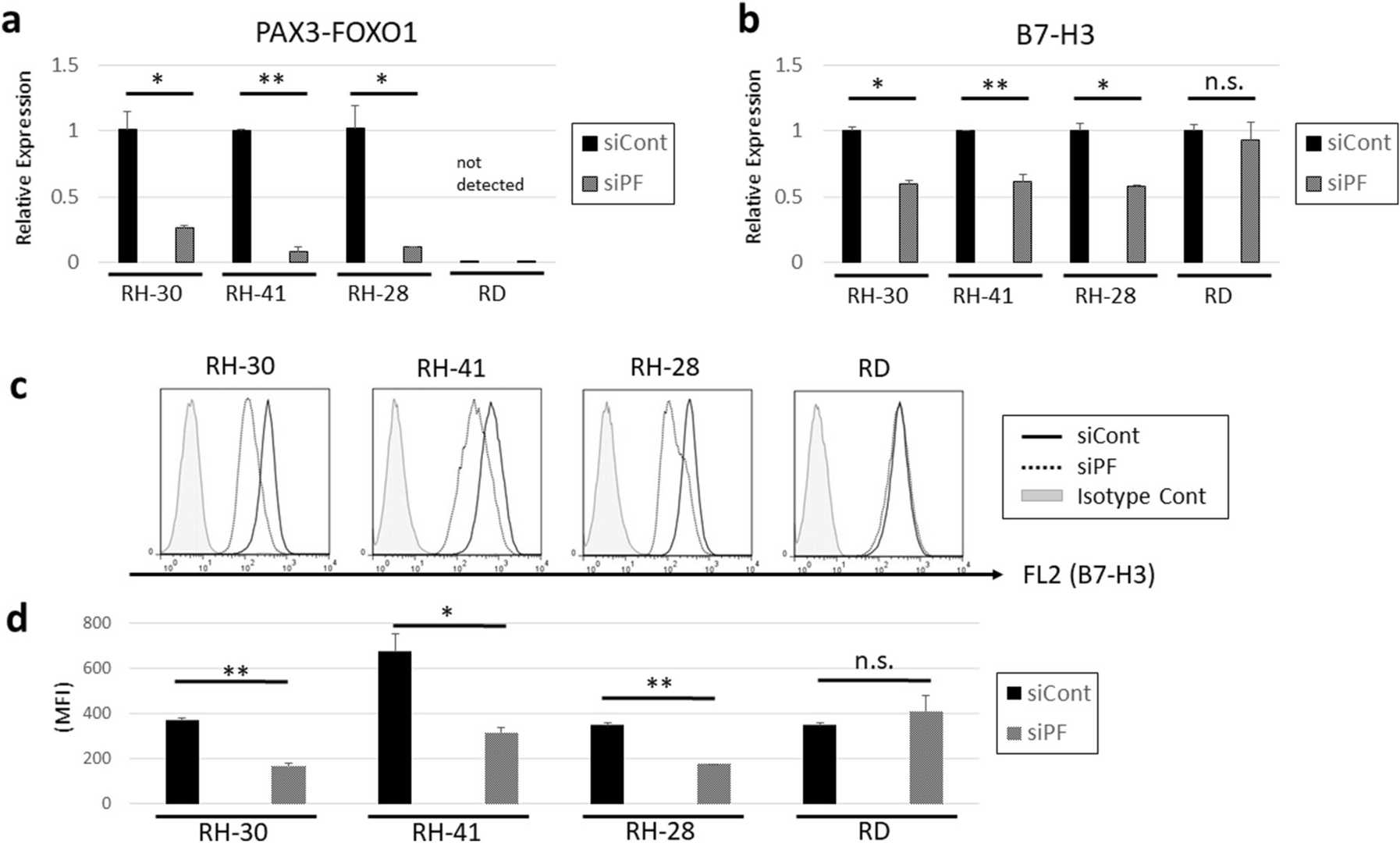 Fig. 1. Knockdown of PAX3-FOXO1 downregulates the expression of B7-H3 (Kanayama, Takuyo, et al. 2021).
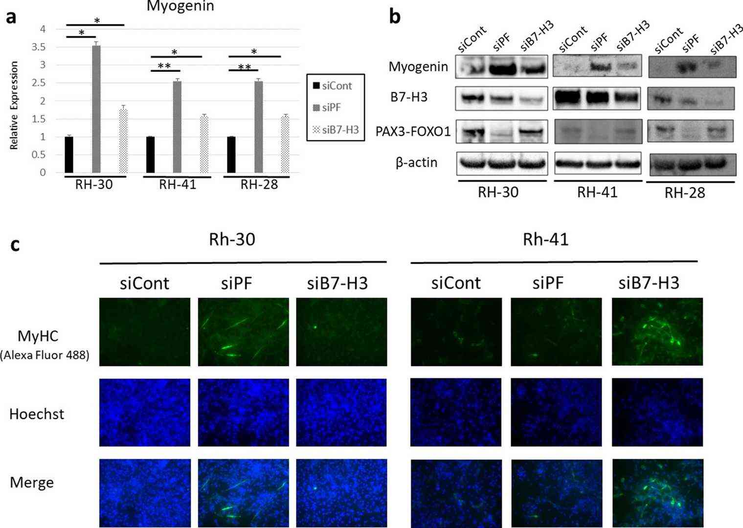 Fig. 2. Knockdown of PAX3-FOXO1 and B7-H3 induces the expression of myogenin and may induce myoblast differentiation (Kanayama, Takuyo, et al. 2021).
A Rhabdomyosarcoma Spheroid Culture Model for In Vitro Evaluation of Hypericin-Based Photodynamic Therapy
Advanced stages of pediatric alveolar rhabdomyosarcoma (RMA) are associated with an unfavorable outcome at established therapeutic strategies, accentuating the need for novel treatment options. Photodynamic therapy (PDT) with hypericin (HYP) has shown strong cytotoxic effects in two-dimensional (2D) cell culture. In order to more accurately mimic in vivo tissue architecture and better predict pharmaceutical response, the aim of this study was to establish a spheroid culture model by which PDT efficacy could be assessed in a three-dimensional (3D) context. Compared to 2D cell culture, a higher treatment resistance was detected, which can be related to spheroid structure and mechanisms of intercellular communication, signal transduction, and gene expression.
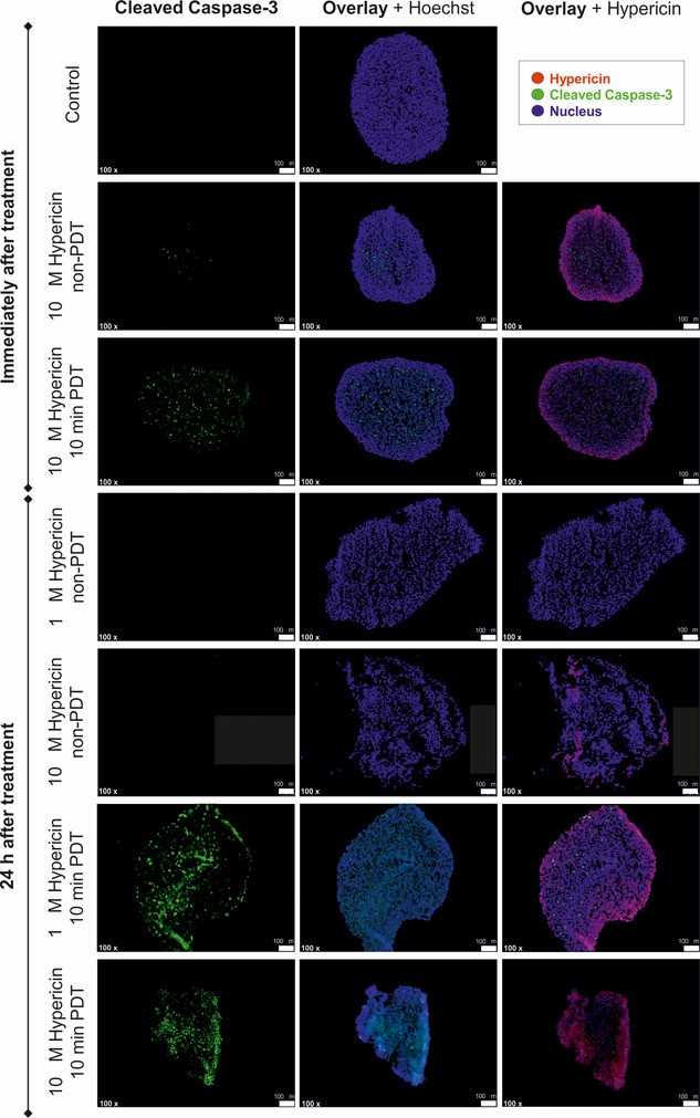 Fig. 3. Evaluation of hypericin (HYP)-based photodynamic therapy (PDT) effects on apoptosis marker expression of RH-30 tumor spheroids (Hempfling, Laura, et al. 2022).
Ask a Question
Write your own review
- You May Also Need

Description: Organism: Homo sapiens (human)Ethnicity: CaucasianAge/Stage: 36 years of ageGender: maleTissue: sarcomaGrowth Properties: monolayerDescription: in vitro established from a primary skin sarcoma (sole ...

Description: Human cell line derived from epithelioid sarcoma. Derived from a different patient from the patient of HS-ES-1 cell line.
Description: Established from an endometrial stromal sarcoma of the uterus of a 76-year-old Caucasian woman; cells were described as demonstrating a mainly mesenchymal phenotype with signs of epithelial ...


Description: Established from the alveololar rhabdomyosarcoma of the liver of a girl after chemotherapy; cells were described to contain a deletion mutation of p53 and to be resistant to FAS- and TRAIL-induced ...

- Adipose Tissue-Derived Stem Cells
- Human Neurons
- Mouse Probe
- Whole Chromosome Painting Probes
- Hepatic Cells
- Renal Cells
- In Vitro ADME Kits
- Tissue Microarray
- Tissue Blocks
- Tissue Sections
- FFPE Cell Pellet
- Probe
- Centromere Probes
- Telomere Probes
- Satellite Enumeration Probes
- Subtelomere Specific Probes
- Bacterial Probes
- ISH/FISH Probes
- Exosome Isolation Kit
- Human Adult Stem Cells
- Mouse Stem Cells
- iPSCs
- Mouse Embryonic Stem Cells
- iPSC Differentiation Kits
- Mesenchymal Stem Cells
- Immortalized Human Cells
- Immortalized Murine Cells
- Cell Immortalization Kit
- Adipose Cells
- Cardiac Cells
- Dermal Cells
- Epidermal Cells
- Peripheral Blood Mononuclear Cells
- Umbilical Cord Cells
- Monkey Primary Cells
- Mouse Primary Cells
- Breast Tumor Cells
- Colorectal Tumor Cells
- Esophageal Tumor Cells
- Lung Tumor Cells
- Leukemia/Lymphoma/Myeloma Cells
- Ovarian Tumor Cells
- Pancreatic Tumor Cells
- Mouse Tumor Cells