
NCTC clone 2472
Cat.No.: CSC-C9559L
Species: Mus musculus (Mouse)
Source: Adipose
Morphology: fibroblast
Culture Properties: monolayer
- Specification
- Background
- Scientific Data
- Q & A
- Customer Review
Strain: C3H/HeN
Tumorigenecity: yes, in C3H/HeN mice
Histocompatibility: H-2k
Histopathology: normal
Note: tested and found negative for ectromelia virus (mousepox)
Shipping Condition: Room Temperature
The NCTC clone 2472 cell line developed from a fibrosarcoma tumor located within the connective tissue of C3H mice. NCTC clone 2472 cells exhibit fibrosarcoma cell characteristics in vitro since their bodies are elongated and slender with spindle or fusiform shapes. Microscopic observation reveals that cell density rises leading to increased cell-to-cell contact and tightly packed cellular clusters. And multicellular structures form when cell density exceeds 100%.
The cell line demonstrates superior syngeneic properties which results in strong compatibility with C3H/He mice. The cell line avoids triggering immune rejection in mice allowing researchers to effectively establish bone cancer models. Mice implanted with this cell line experience bone destruction and pain which creates a standard bone cancer pain model. Scientists often use this model to explore how bone cancer pain functions through neurotransmitter release, astrocyte activation and inflammatory factor expression. Studies demonstrate that the tumor area shows significant elevation of CCL2 (monocyte chemoattractant protein-1) released by NCTC clone 2472 cells which contributes to thermal and mechanical hyperalgesia development.
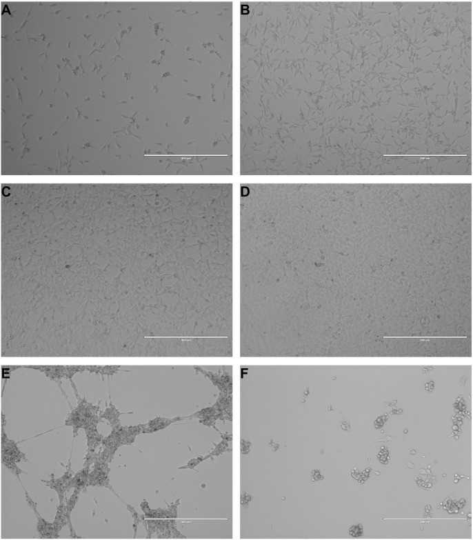 Fig. 1. Images are representative of NCTC-2472 fibrosarcoma cells at increasing levels of dish confluency at 20% confluency (a), 50% confluency (b), 80% confluency (c), and 100% confluency (d). Upon surpassing 100% confluency, cells began to clump together into large, multi-cellular strands (e). While NCTC-2472 are robust, lack of fresh nutrients will induce a poor health state (f) with observed cellular shrinkage, blebbing, and loss of adherence (Ortiz YT, Shamir LG, et al., 2024).
Characterization of Commercially Available Murine Fibrosarcoma NCTC-2472 Cells
Cancer-induced bone pain (CIBP) is a severe neuropathic pain caused by tumor metastasis to bone, significantly impacting patients' quality of life. Current treatments focus on symptom management, but novel therapeutics are needed. The NCTC-2472 murine fibrosarcoma cell line is commonly used in preclinical CIBP models. This study characterized NCTC-2472 cells both in vitro and in vivo to assess their viability and suitability for evaluating new anti-cancer and analgesic compounds.
NCTC-2472 fibrosarcoma cells did not display prominent morphological markers indicative of apoptosis in regular culture. Throughout continued cell passaging, NCTC-2472 cells exhibited typical cellular morphology associated with fibroblast type cells that include branched cytoplasmic extensions and a general spindle shape body pattern with bipolar presentation (Fig 1a). There was no observed shrinking of the nucleus, no cellular blebbing, minimal presence of cellular debris, and no loss of adherence in low passage numbers (Fig 1b), moderate passage numbers (Fig 1c), or in high passage numbers (Fig 1d) amongst any cells. To determine the self-renewal capacity of NCTC-2472 fibroblasts, we measured and calculated the proliferative ability of the cells via CPDL measurements (Fig 1e). A consistently linear increase in the rate of cell proliferation was observed and indicated that despite prolonged passaging, there was no impact on cellular proliferation.
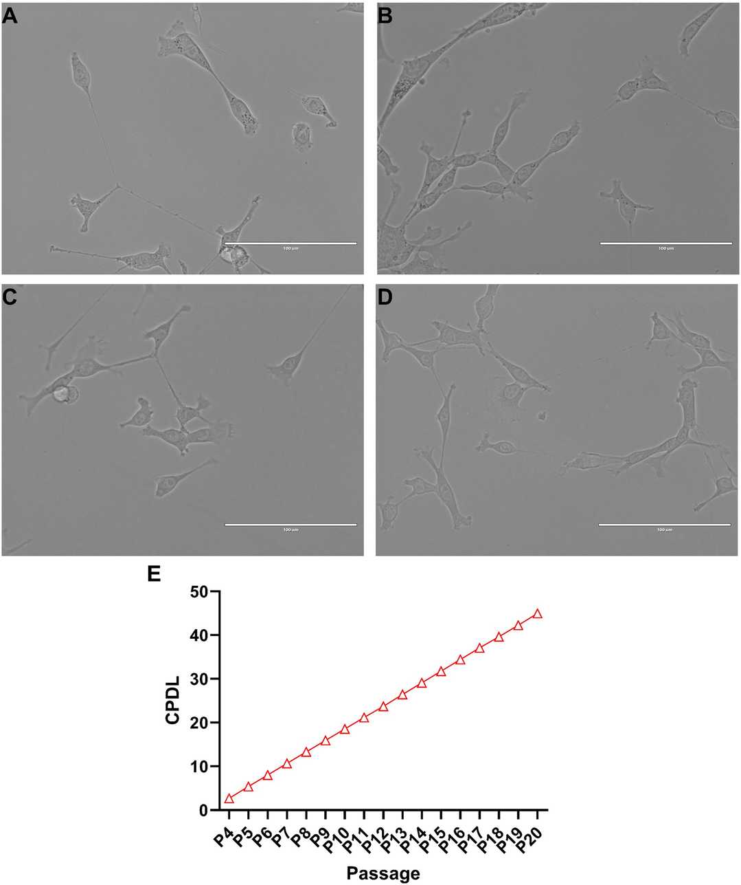 Fig. 1. Culture of NCTC-2472 fibrosarcoma cells and identification of cumulative population doubling level (CPDL) (Ortiz YT, Shamir LG, et al., 2024).
LPA Signaling Contributed to Hypersensitivity Produced by Bone Cancer
Bone cancer pain is a severe condition affecting over 60% of patients, with current treatments often being insufficient. The mechanisms involve interactions between cancer cells and nociceptive neurons, with autotaxin (ATX) and lysophosphatidic acid (LPA) playing key roles in cancer progression and pain signaling. This study investigates the role of ATX–LPA–LPAR signaling in cancer pain using in vitro and in vivo methods.
Consistent with prior studies mechanical and heat hypersensitivity developed within 10 days after implanting mouse NCTC 2472 fibrosarcoma cells into the calcaneus bone of male and female mice, with no sex differences. Implanting nonmalignant NCTC 929 fibroblasts caused no tumors or hypersensitivity in naive mice. Tumor-bearing (TB) mice showed elevated serum LPA levels (Fig. 2A), specifically 18:1-, 18:2-, and 20:4-LPA (Fig. 2B), while 16:0- and 22:6-LPA remained unchanged. LPA levels were not measured in rare non-hypersensitive mice. In vitro, 18:1-LPA (10 µM, 24h) promoted fibrosarcoma cell proliferation (Fig. 2C), consistent with prior protocols. Given elevated LPA and its LPAR1,3,5 targeting in DRG, LPAR antagonist H2L5765834 (5 mg/kg, intraplantar) reduced mechanical and heat hypersensitivity in TB mice versus vehicle (Figs. 2D, E). Lower doses (1 mg/kg) or contralateral administration had no effect, indicating localized action.
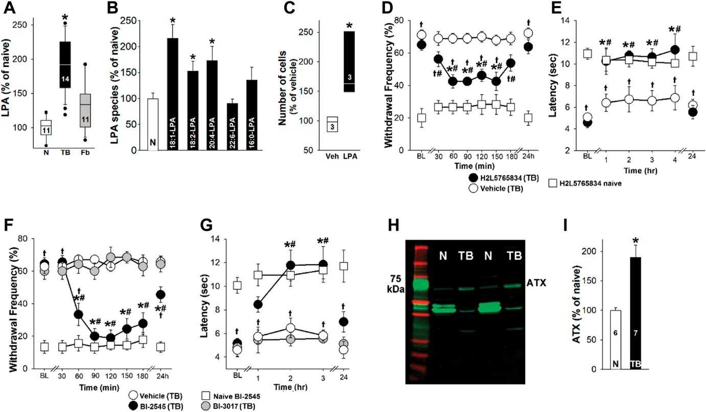 Fig. 2. Lysophosphatidic acid signaling contributed to hypersensitivity produced by bone cancer (Iryna A K, Sergey G K, et al., 2023).
Ask a Question
Write your own review

Description: Described as secreting a mouse monoclonal antibody (IgG2a) detecting all fibers in skeletal muscle and myosin heavy chains on Western blots and detecting mammalian, chicken, zebrafish, axolotl, ...

Description: Animals were immunized with the B6.1 mouse cytotoxic T cell line.


Description: neuroglial and neuronal character coexpressing ependymoma cell line.

Description: Established by irradiation of the adherent cells in long-term bone marrow cultures derived from C3H/HeNSlc strain mice
- Adipose Tissue-Derived Stem Cells
- Human Neurons
- Mouse Probe
- Whole Chromosome Painting Probes
- Hepatic Cells
- Renal Cells
- In Vitro ADME Kits
- Tissue Microarray
- Tissue Blocks
- Tissue Sections
- FFPE Cell Pellet
- Probe
- Centromere Probes
- Telomere Probes
- Satellite Enumeration Probes
- Subtelomere Specific Probes
- Bacterial Probes
- ISH/FISH Probes
- Exosome Isolation Kit
- Human Adult Stem Cells
- Mouse Stem Cells
- iPSCs
- Mouse Embryonic Stem Cells
- iPSC Differentiation Kits
- Mesenchymal Stem Cells
- Immortalized Human Cells
- Immortalized Murine Cells
- Cell Immortalization Kit
- Adipose Cells
- Cardiac Cells
- Dermal Cells
- Epidermal Cells
- Peripheral Blood Mononuclear Cells
- Umbilical Cord Cells
- Monkey Primary Cells
- Mouse Primary Cells
- Breast Tumor Cells
- Colorectal Tumor Cells
- Esophageal Tumor Cells
- Lung Tumor Cells
- Leukemia/Lymphoma/Myeloma Cells
- Ovarian Tumor Cells
- Pancreatic Tumor Cells
- Mouse Tumor Cells