- Specification
- Background
- Scientific Data
- Q & A
- Customer Review
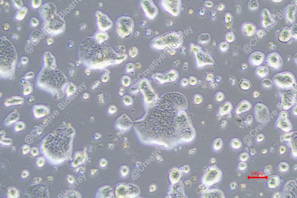
The HT55 cell line originates from human colorectal adenocarcinoma tissue and was first established in 1977, as described by Watkins, Sanger, and others. Throughout more than 120 passages this cell line preserved its highly differentiated state and did not develop undifferentiated derivatives. The HT55 cell line displays diverse cell shapes while retaining epithelial characteristics. This cell line shows an extended G1 phase and S phase in its growth characteristics because its differentiated state requires complex macromolecular synthesis. Colon adenocarcinoma growth cycles frequently display extended S phases. In athymic mice, the HT55 cell line can induce ossification and stimulate the formation of transient nodules in CBA mouse embryonic fibroblasts. These characteristics suggest that the HT55 cell line possesses a certain tumorigenic potential in vivo.
The HT55 cell line has a wide range of applications in scientific research. Firstly, it is used to study the molecular mechanisms of colorectal cancer, including its growth cycle, ossification capacity, and RNA content production. In cancer treatment research, the HT55 cell line is utilized to evaluate the efficacy of various drugs.
Exploration of Predictive Marker Proteins for Sensitivity to Anti‑EGFR Monoclonal Antibodies in Colorectal Cancer Cell Lines
Proteins from cancer cells are increasingly studied as biomarkers for chemotherapy efficacy. Nagamine et al. identified a sensitive biomarker for anti-EGFR monoclonal antibodies (anti-EGFR mAbs) in colorectal cancer (CRC) cell lines. Using mass spectrometry, proteins from six CRC cell lines with varying cetuximab sensitivity were analyzed.
Six CRC cell lines with varying cetuximab sensitivity were used: CACO2, C10, HT55, C99, SW48, and COLO320DM. They were divided into three groups: Cmab-S (C99, SW48), Cmab-PR (C10, HT55), and Cmab-R. PCA was performed on MS data from Cmab-R and Cmab-S using LC-TOF MS. A total of 1,599 and 1,012 cytoplasmic protein peaks, and 2,370 and 3,478 membrane protein peaks were detected for Cmab-R and Cmab-S, respectively. After filtering, 148 and 146 cytoplasmic protein peaks, and 363 and 267 membrane protein peaks were identified as significantly correlated with cetuximab sensitivity. Proteins were identified by MS fit analysis, with cytoplasmic LDHB showing the highest MOWSE score (Fig. 1).
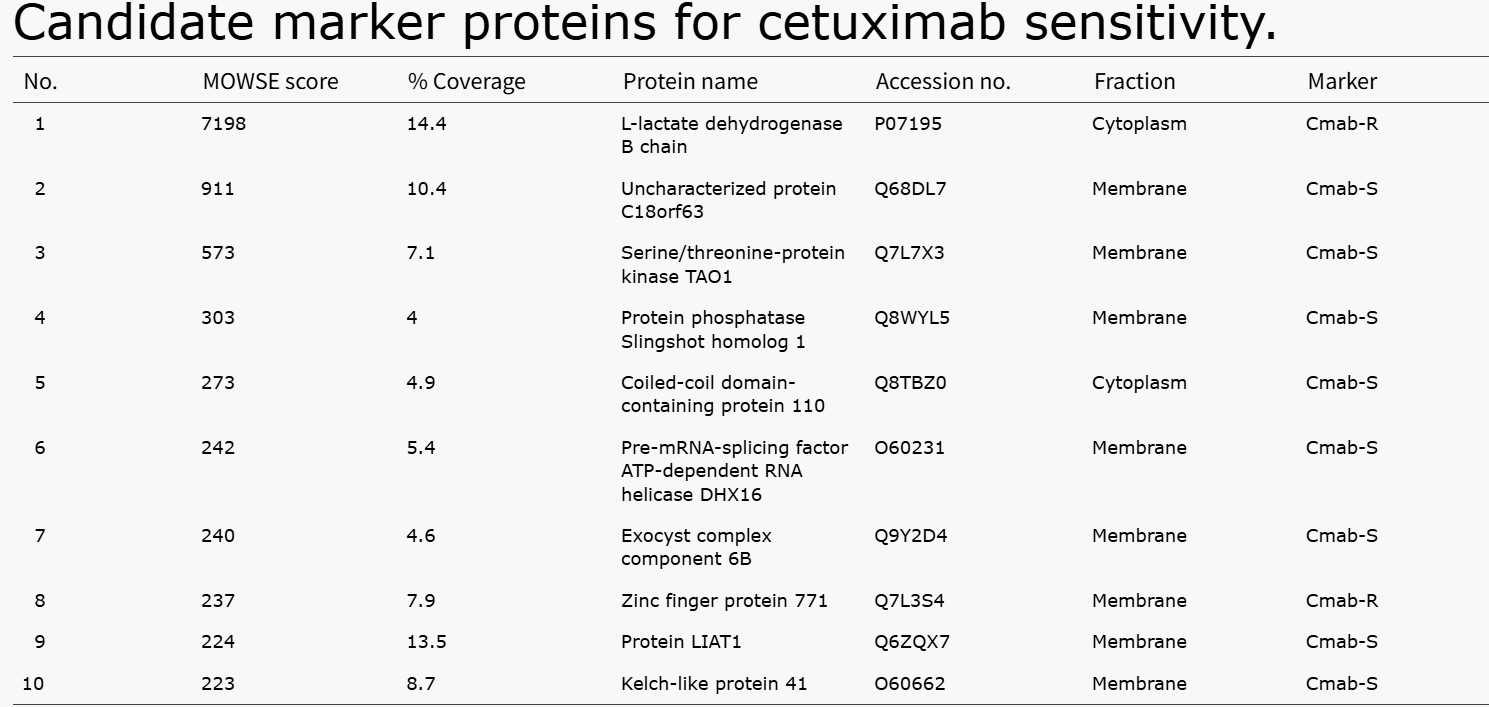 Fig. 1. Candidate marker proteins for cetuximab sensitivity (Nagamine A, Araki T, et al., 2019).
LY6G6D-TDB is Active Against Colorectal Cancer Cell Lines with a Broad Range of Target Expression
Colorectal cancer is a major global health issue with poor survival rates, especially for metastatic cases. Immune checkpoint inhibitors (ICIs) have shown efficacy in mismatch-repair–deficient or microsatellite instability–high (MSI-H) colorectal cancer, but most patients with microsatellite-stable (MSS) or MSI-low cancer do not benefit. Wang's team developed a novel T-cell–dependent bispecific antibody (LY6G6D-TDB) targeting the tumor-associated antigen LY6G6D for treating colorectal cancer.
They evaluated the correlation between LY6G6D expression level and LY6G6D-TDB potency by selecting four colorectal cancer cell lines (COLO320DM, GPD20, HT55 and LS1034) with surface LY6G6D ranging from 500 to 10,000 binding sites per cell. IHC was also used to determine LY6G6D expression in these cell lines, and the results of the two methods correlated well (Fig. 2A and B). The levels of LY6G6D in these cell lines correspond to those in colorectal cancer tumors having low to high levels of LY6G6D. Regardless of LY6G6D expression level, the four colorectal cancer cell lines showed comparable sensitivity to 1G4/CD3H–mediated cytotoxicity with EC50 values ranging from 7.11 to 19.49 ng/mL. No cell killing was observed in the LY6G6D-negative HEK 293 cells (Fig. 2C). These data suggest that target cell killing by LY6G6D-TDB is not strictly driven by the level of LY6G6D, and LY6G6D-TDB can mediate killing of colorectal cancer cell lines with a wide range of LY6G6D expression, including cell lines with low levels of the target.
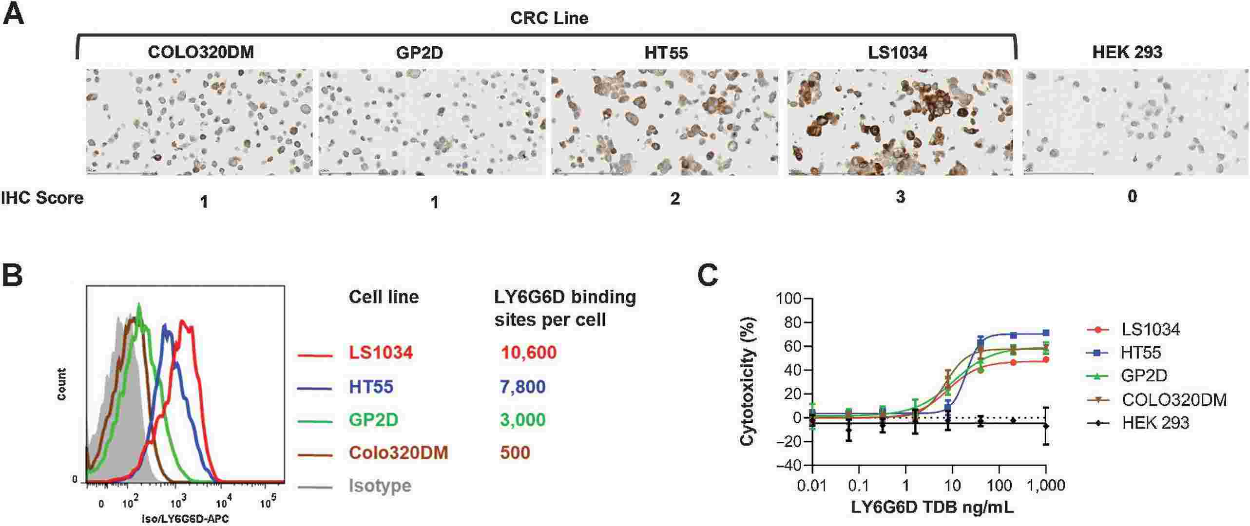 Fig. 2. LY6G6D-TDB is active against colorectal cancer cell lines with a broad range of LY6G6D level (Wang P, Sun LL, et al., 2022).
Ask a Question
Write your own review

Description: Established from an adenocarcinoma located in the ascending colon of a 68 year-old male patient. The adenocarcinoma was well differentiated and determined to be Dukes' stage B. Imperial College ...

Description: This is one cell line out of a series of colon carcinoma cell lines established by PD Dr. Michael Linnebacher.
Description: Rectal carcinoma from a Japanese patient. Cell growth is slow.
Description: The C80 cell line was established from a 69-year old male patient with a moderately well differentiated adenocarcinoma of the rectum classified as Dukes' stage D.
Description: RKO is a poorly differentiated colon carcinoma cell line developed by Michael Brattain.
- Adipose Tissue-Derived Stem Cells
- Human Neurons
- Mouse Probe
- Whole Chromosome Painting Probes
- Hepatic Cells
- Renal Cells
- In Vitro ADME Kits
- Tissue Microarray
- Tissue Blocks
- Tissue Sections
- FFPE Cell Pellet
- Probe
- Centromere Probes
- Telomere Probes
- Satellite Enumeration Probes
- Subtelomere Specific Probes
- Bacterial Probes
- ISH/FISH Probes
- Exosome Isolation Kit
- Human Adult Stem Cells
- Mouse Stem Cells
- iPSCs
- Mouse Embryonic Stem Cells
- iPSC Differentiation Kits
- Mesenchymal Stem Cells
- Immortalized Human Cells
- Immortalized Murine Cells
- Cell Immortalization Kit
- Adipose Cells
- Cardiac Cells
- Dermal Cells
- Epidermal Cells
- Peripheral Blood Mononuclear Cells
- Umbilical Cord Cells
- Monkey Primary Cells
- Mouse Primary Cells
- Breast Tumor Cells
- Colorectal Tumor Cells
- Esophageal Tumor Cells
- Lung Tumor Cells
- Leukemia/Lymphoma/Myeloma Cells
- Ovarian Tumor Cells
- Pancreatic Tumor Cells
- Mouse Tumor Cells