
Caov-3
Cat.No.: CSC-C9094W
Species: Homo sapiens (Human)
Source: Ovary
Morphology: epithelial
- Specification
- Background
- Scientific Data
- Q & A
- Customer Review
Caov-3 cells are an invaluable resource in biological science, specifically for the study of ovarian cancer. Derived from the ovary of a 54-year-old Caucasian female with adenocarcinoma, these cells provide researchers with a representative model of high-grade ovarian cancer. The cell line was established in 1976 and has since become a widely used tool in numerous studies, achieving an impressive track record with 614 product citations.
With their epithelial morphology, Caov-3 cells closely resemble the characteristics of primary ovarian cancer cells. When cultured, these cells form tightly packed colonies that mimic the behavior observed in the human body. Their unique properties make them an ideal choice for researchers investigating the growth, behavior, and response of ovarian carcinoma cells. Furthermore, Caov-3 cells express multiple tumor-associated antigens, including NB/70K, CA-125, Ba-2, and Ca-1. These antigens are key markers used to identify and characterize ovarian cancer cells. Their presence in the Caov-3 cell line enhances its utility in targeted therapies and immunotherapies studies. The genetic makeup of Caov-3 cells reveals significant abnormalities, further contribute to their relevance in ovarian cancer research. These cells harbor a nonsense mutation in the p53 gene, a tumor suppressor gene frequently implicated in various cancers. Additionally, they possess multiple copies of the ovarian cancer oncogene PIK3CA, which plays a crucial role in cancer development and progression. Such genetic aberrations allow researchers to study the effects of these mutations on ovarian cancer biology and explore potential therapeutic targets.
Regarding drug sensitivity, Caov-3 cells exhibit responsiveness to several commonly used chemotherapeutic agents. Vinblastine, cisplatin, and adriamycin have been shown to elicit a notable effect on these cells. This sensitivity allows researchers to evaluate the efficacy of different drugs, develop new treatment strategies, and study drug resistance mechanisms. Another feature of Caov-3 cells is their behavior in different culture conditions. While these cells fail to grow in soft agar, they demonstrate tumorigenicity when injected into immunocompromised mice. This observation highlights the relevance of the Caov-3 cell line in vivo to study and explore cancer progression and metastasis. In addition to their numerous applications in research, Caov-3 cells are particularly suitable for 3D cell culture experiments. Their epithelial morphology and ability to form tight colonies make them ideal for studying cell-cell interactions, tissue organization, and the behavior of ovarian cancer cells in a more physiologically relevant environment.
The Expression of CHD1L is Increased in Ovarian Cancer Tissues and Cell Lines
TNMPLOT database was used to analyzed chromodomain helicase/ATPase DNA-binding protein 1-like gene (CHDIL) expression in ovarian cancer (OC) tissues and normal ovarian tissues. As shown in Fig. 1A, CHD1L expression was notably upregulated in OC tissues compared with that in the normal ovarian tissues. RT-qPCR and Western blot were used to detect the expression of CHD1L in OC cell lines, and the results showed that the expression of CHD1L in OC cell lines was significantly increased compared with IOSE80 cells (Fig. 1B and C). Among them, CHD1L had the highest expression in CAOV-3 cells.
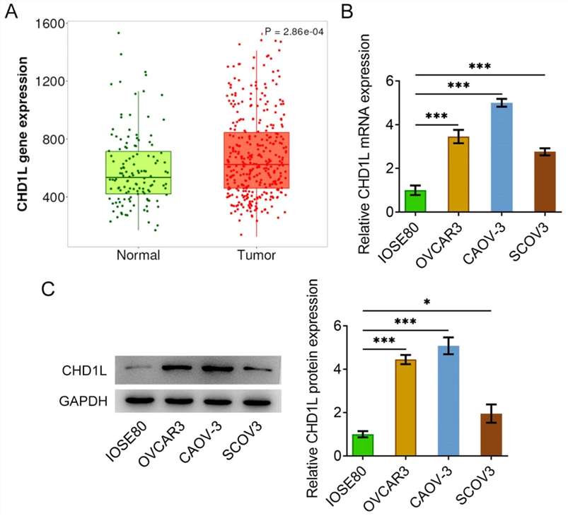 Fig. 1. The expression of CHD1L was increased in OC tissues and cell lines (Qiao, K., Guan, Y. and Xing, W., 2025).
Exposure to FSS Decreases Sensitivity to BPD-PDT
Malignant ascites, or excessive fluid accumulation in the peritoneum, is a common occurrence in patients with high-grade serous ovarian carcinoma (HGSOC). There is a growing interest in determining the effects of ascitic fluid streams, and the associated fluid shear stress (FSS), on ovarian cancer cell phenotypes, migration, and therapy response. While it is crucial to investigate the effects of FSS on phenotypic changes and chemotherapy response in HGSOC, chemotherapy alone is unlikely to produce meaningful breakthroughs in the management of this disease. Photodynamic therapy (PDT) is a mechanistically distinct modality, which relies on a photochemistry-based generation of reactive molecular species (RMS) within a confined area of irradiation. In addition to its direct RMS-mediated cytotoxicity, PDT has been shown to synergistically enhance Pt-based chemotherapy in the in vitro and in vivo models for ovarian carcinomatosis.
The present study employed benzoporphyrin derivative (BPD), a clinical photosensitizer known for its ability to synergize with Pt-based chemotherapies. BPD-PDT was investigated as a standalone modality, as well as in combination with cisplatin and doxorubicin as a means of overcoming FSS-induced Pt resistance in an in vitro perfusion model. In a static culture, OVCAR-3 and Caov-3 cells showed similar sensitivity to BPD-PDT (Fig. 2B). FSS exposure, however, decreased sensitivity to BPD-PDT in both cell lines (Fig. 2C). In all instances, OVCAR-3 and Caov-3 cells exposed to FSS showed significant resistance to BPD-PDT at low energy densities (0.1-0.5?J/cm2).
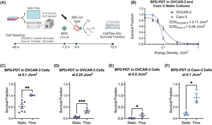 Fig. 2. BPD-PDT dose-responses in OVCAR-3 and Caov-3 cells under static and flow conditions (Overchuk, Marta, et al. 2024).
Ask a Question
Write your own review
Description: established from ovary of a 47-year-old female with ovarian endometrioid adenocarcinoma

Description: The A2780 human ovarian cancer cell line was established from tumour tissue from an untreated patient. Cells grow as a monolayer and in suspension in spinner cultures. A2780 is the parent line to the ...
Description: Japanese ovarian serous adenocarcinoma. Said CA125 producing. Cell growth is slow.

Description: Established post-hysterectomy from the tumor tissue of a 65-year-old Japanese woman with malignant ovarian carcinoma (stage: FIGO IIIc; histology: poorly differentiated serous papillary ...

- Adipose Tissue-Derived Stem Cells
- Human Neurons
- Mouse Probe
- Whole Chromosome Painting Probes
- Hepatic Cells
- Renal Cells
- In Vitro ADME Kits
- Tissue Microarray
- Tissue Blocks
- Tissue Sections
- FFPE Cell Pellet
- Probe
- Centromere Probes
- Telomere Probes
- Satellite Enumeration Probes
- Subtelomere Specific Probes
- Bacterial Probes
- ISH/FISH Probes
- Exosome Isolation Kit
- Human Adult Stem Cells
- Mouse Stem Cells
- iPSCs
- Mouse Embryonic Stem Cells
- iPSC Differentiation Kits
- Mesenchymal Stem Cells
- Immortalized Human Cells
- Immortalized Murine Cells
- Cell Immortalization Kit
- Adipose Cells
- Cardiac Cells
- Dermal Cells
- Epidermal Cells
- Peripheral Blood Mononuclear Cells
- Umbilical Cord Cells
- Monkey Primary Cells
- Mouse Primary Cells
- Breast Tumor Cells
- Colorectal Tumor Cells
- Esophageal Tumor Cells
- Lung Tumor Cells
- Leukemia/Lymphoma/Myeloma Cells
- Ovarian Tumor Cells
- Pancreatic Tumor Cells
- Mouse Tumor Cells