
- Specification
- Background
- Scientific Data
- Q & A
- Customer Review
OACP4 C is a human esophageal adenocarcinoma cell line established from a primary adenocarcinoma located in the gastric cardia of a 55-year-old Caucasian male patient with lymph node involvement and pleural metastases. The cell line exhibits adherent growth and displays pleiomorphic morphology, containing both epithelial-like and fibroblastoid cells with prominent cytoplasmic protrusions. OACP4 C has been shown to be tumorigenic in immunodeficient mouse models, including NMRI nude and NOD-SCID mice, making it a valuable in vitro and in vivo model for upper gastrointestinal cancer research.
Genetic characterization of OACP4 C has identified multiple molecular alterations associated with esophageal adenocarcinoma, including a homozygous TP53 mutation (c.574C>T, p.Gln192Ter). Whole-genome and transcriptomic analyses have further confirmed its relevance as a representative model of esophageal and gastric cardia adenocarcinoma. OACP4 C is widely used in studies of tumor progression, genomic instability, drug sensitivity, signaling pathway analysis, and biomarker discovery in gastroesophageal cancers. Its stable growth characteristics and well-characterized genomic background make it a useful platform for translational oncology research and preclinical therapeutic evaluation.
Intrinsic Therapy Resistance in OAC Cell Lines
Oesophageal adenocarcinoma (OAC) patients often have poor prognoses due to inherent resistance to chemoradiotherapy, though the molecular mechanisms remain unclear. MicroRNAs (miRNAs) are potential biomarkers for treatment response. Butz et al. aimed to identify specific miRNAs associated with intrinsic resistance in OAC.
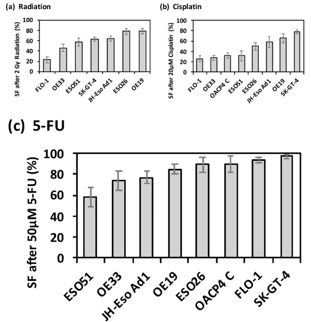
Clonogenic assays assessed baseline radiosensitivity across eight cell lines. Seven lines showed consistent resistance to 2 Gy radiation and were retained for analysis; OACP4C exhibited variable responses and was excluded (Fig. 1a). FLO-1 was the most sensitive (survival fraction [SF] = 23% ± 5.4%), while ESO26 and OE19 were the most resistant (SF = 78% ± 6.1% and 79% ± 5.1%; Fig. 1a).
Apoptosis assays evaluated intrinsic chemoresistance. For cisplatin, SFs ranged from 26% ± 3.6% (FLO-1, most sensitive) to 78% ± 3.9% (SK-GT-4, most resistant) (Fig. 1b). OE33, OACP4C, and ESO51 also showed low cisplatin resistance (SF ≈ 28-32%). For 5-FU, five of eight lines (OE19, ESO26, OACP4C, FLO-1, SK-GT-4) exhibited high resistance (SF > 85%), while ESO51 was the most sensitive (SF = 58% ± 3.6%; Fig. 1c). These data establish a panel of OAC cell lines with defined resistance phenotypes for biomarker discovery.
Ask a Question
Write your own review


Description: Rhabdoid tumor of kidney (formerly classified as Wilms' tumor)


Description: The cells express the transforming gene of adenovirus 5. Adenovirus 5 DNA from both the right and left ends of the viral genome are present. The line is excellent for titrating human adenoviruses. ...
- Adipose Tissue-Derived Stem Cells
- Human Neurons
- Mouse Probe
- Whole Chromosome Painting Probes
- Hepatic Cells
- Renal Cells
- In Vitro ADME Kits
- Tissue Microarray
- Tissue Blocks
- Tissue Sections
- FFPE Cell Pellet
- Probe
- Centromere Probes
- Telomere Probes
- Satellite Enumeration Probes
- Subtelomere Specific Probes
- Bacterial Probes
- ISH/FISH Probes
- Exosome Isolation Kit
- Human Adult Stem Cells
- Mouse Stem Cells
- iPSCs
- Mouse Embryonic Stem Cells
- iPSC Differentiation Kits
- Mesenchymal Stem Cells
- Immortalized Human Cells
- Immortalized Murine Cells
- Cell Immortalization Kit
- Adipose Cells
- Cardiac Cells
- Dermal Cells
- Epidermal Cells
- Peripheral Blood Mononuclear Cells
- Umbilical Cord Cells
- Monkey Primary Cells
- Mouse Primary Cells
- Breast Tumor Cells
- Colorectal Tumor Cells
- Esophageal Tumor Cells
- Lung Tumor Cells
- Leukemia/Lymphoma/Myeloma Cells
- Ovarian Tumor Cells
- Pancreatic Tumor Cells
- Mouse Tumor Cells