
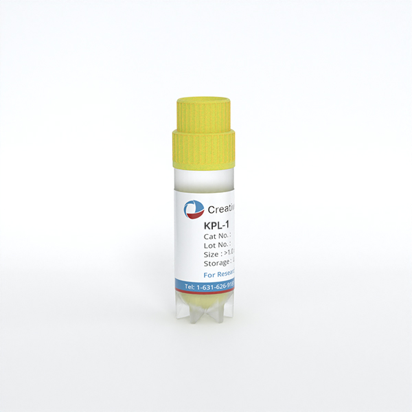
KPL-1
Cat.No.: CSC-C0389
Species: Homo sapiens (Human)
Source: Pleural Effusion Metastasis
Morphology: adherent growing in monolayers (very dense colonies)
Culture Properties: monolayer
- Specification
- Background
- Scientific Data
- Q & A
- Customer Review
Immunology: cytokeratin +, cytokeratin-7 -, cytokeratin-8 +, cytokeratin-17 -, cytokeratin-18 +, cytokeratin-19 +, desmin -, endothel -, EpCAM +, GFAP -, neurofilament -
KPL-1 is an authenticated human breast adenocarcinoma cell line established in 1995 from the malignant effusion of a patient with invasive ductal carcinoma. Classified within the luminal A molecular subtype, KPL-1 is estrogen receptor (ER)-positive, progesterone receptor (PR)-negative, and HER2-negative, with no amplification of c-erbB-2, c-myc, H-ras, or N-ras. Notably, it exhibits androgen receptor (AR) expression and differs from its parental MCF-7 background in MUC-1 and AR profiles, offering a distinct luminal model for comparative studies.
A defining advantage of KPL-1 is its estrogen-independent proliferation in vitro, making it particularly valuable for dissecting hormone-refractory mechanisms within an ER-positive context. The cells robustly express epithelial markers including cytokeratin, carcinoembryonic antigen, and CA 15-3, and secrete high levels of tissue polypeptide antigen (TPA) in a growth-phase-dependent manner—providing a quantifiable biomarker system that correlates directly with tumor burden in vivo.
KPL-1 demonstrates exceptional tumorigenicity, forming rapidly growing xenografts in female athymic nude mice with documented regional axillary lymph node metastasis. Its hypertriploid karyotype, authenticated STR profile, and extensive characterization across transcriptomic and pharmacogenomic platforms ensure experimental reproducibility. With its authenticated origin, defined luminal phenotype, hormone-independent growth capability, and proven in vivo metastatic potential, KPL-1 stands as a robust and versatile platform for investigating breast cancer biology, tumor-associated antigen kinetics, and therapeutic response.
Non-Enzymatic SETD1A Activity Drives Breast Cancer Cell Proliferation
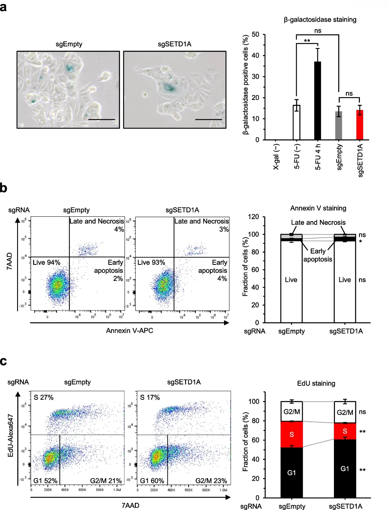
Phenotype changes caused by SETD1A deletion varies across different carcinomas and cell lines. To determine the KPL-1 breast cancer cell phenotype induced by SETD1A knockout, we assessed senescence, apoptosis and cell-cycle arrest at seven days post-Dox treatment, using sgRNA-expressing iCas9 KPL-1 cells.
SETD1A knockout did not affect the percentage of β-galactosidase-positive cells (Fig. 1a). This finding suggests that the loss of SETD1A suppresses cell proliferation through alternative mechanisms in the KPL-1 cell line. While SETD1A knockout increased the proportion of early apoptotic cells, the effect was modest and insufficient to account for the significant impact observed in the cell growth assay seven days post-Dox treatment (Fig. 1b). SETD1A knockout significantly increased the proportion of cells in the G1 phase while reducing the proportion in the S phase (Fig. 1c). These results indicate that SETD1A deletion primarily induces cell cycle arrest at the G1 phase in KPL-1 breast cancer cells.
Ask a Question
Write your own review
- You May Also Need
Description: MCF10DCIS.com is a clonal breast cancer cell line derived from a xenograft originating from premalignant MCF10AT cells that were injected into severe combined immune-deficient mice. MCF10AT is ...
Description: Species: human, Caucasian female 69 years old; Tissue: breast; Tumor: adenocarcinoma; Transfected with ER-pBact, human estrogen receptor cDNA, under control of the human beta actin promotor, ...
Description: Histopathology: breast cancerNote: NK (natural killer) cell resistant clone
Description: ZR-75-30 was derived from malignant ascites fluid from a 47-year-old premenopausal Black woman with infiltrating ductal carcinoma.
Description: Species: human - female, 74 years old, CaucasianTumorigenecity: yes, in nude miceIsoenzyme: G6PD, B; PGM3, 1; PGM1, 1; ES-D, 1; Me-2, 0; AK-1, 1; GLO-1, 1Histopathology: carcinoma, ductal
Description: Species: human, Caucasian female 69 years old; Tissue: breast; Tumor: adenocarcinoma; Transfected with the exon 5 defective variant of human estrogen receptor cDNA, cloned in pZeoSV vector, resistant ...
- Adipose Tissue-Derived Stem Cells
- Human Neurons
- Mouse Probe
- Whole Chromosome Painting Probes
- Hepatic Cells
- Renal Cells
- In Vitro ADME Kits
- Tissue Microarray
- Tissue Blocks
- Tissue Sections
- FFPE Cell Pellet
- Probe
- Centromere Probes
- Telomere Probes
- Satellite Enumeration Probes
- Subtelomere Specific Probes
- Bacterial Probes
- ISH/FISH Probes
- Exosome Isolation Kit
- Human Adult Stem Cells
- Mouse Stem Cells
- iPSCs
- Mouse Embryonic Stem Cells
- iPSC Differentiation Kits
- Mesenchymal Stem Cells
- Immortalized Human Cells
- Immortalized Murine Cells
- Cell Immortalization Kit
- Adipose Cells
- Cardiac Cells
- Dermal Cells
- Epidermal Cells
- Peripheral Blood Mononuclear Cells
- Umbilical Cord Cells
- Monkey Primary Cells
- Mouse Primary Cells
- Breast Tumor Cells
- Colorectal Tumor Cells
- Esophageal Tumor Cells
- Lung Tumor Cells
- Leukemia/Lymphoma/Myeloma Cells
- Ovarian Tumor Cells
- Pancreatic Tumor Cells
- Mouse Tumor Cells