

KHYG-1
Cat.No.: CSC-C0784
Species: Homo sapiens (Human)
Source: Blood; Peripheral Blood
Morphology: round to polymorph cells growing singly and in clumps in suspension
Culture Properties: suspension
- Specification
- Background
- Scientific Data
- Q & A
- Customer Review
- Documents
CSF1PO 12
D5S818 9,11
D7S820 10,11
D13S317 9,13
D16S539 9
TH01 6
TPOX 9,11
vWA 17
b. The study of enhanced cytotoxicity by NK cells
c. Cell-Based assay
d. In vivo efficacy study
Viruses: ELISA: reverse transcriptase negative; PCR: EBV -, HBV -, HCV -, HHV-8 -, HIV-1 -, HIV-2 -, HTLV-I/II -, MLV -, SMRV -
Store in liquid nitrogen.
Biosafety classification is based on U.S. Public Health Service Guidelines, it is the responsibility of the customer to ensure that their facilities comply with biosafety regulations for their own country.
KHYG-1 cells are a cell line derived from the peripheral blood of a 45-year-old woman diagnosed with aggressive natural killer (NK) cell leukemia. They were established in 1997 and have since been widely used in research related to NK cell biology, cancer, immunology, and cytotoxicity studies.
The primary feature that distinguishes KHYG-1 cells is their potent cytotoxic activity, which is a characteristic trait of NK cells. NK cells are a type of lymphocyte in the immune system that plays a crucial role in recognizing and eliminating infected or cancerous cells. KHYG-1 cells retain this cytotoxic potential even after being established as a cell line. This property makes them a valuable tool for studying NK cell biology, their mechanisms of cytotoxicity, and their interactions with target cells.
Another notable aspect of KHYG-1 cells is the presence of a point mutation in exon 7 of the TP53 (p53) gene. The p53 gene encodes a tumor suppressor protein that plays a critical role in regulating cell division and preventing the formation of cancerous cells. The mutation in KHYG-1 cells suggests an alteration in the function of the p53 protein, which may have implications for studying the role of p53 in NK cell leukemia and related diseases.
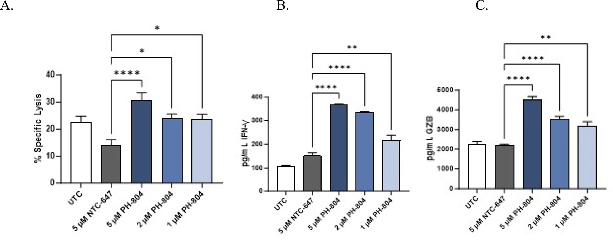
INTASYL self-delivering RNAi decreases TIGIT expression, enhancing NK cell cytotoxicity: a potential application to increase the efficacy of NK adoptive cell therapy against cancer
Background
Natural killer (NK) cells are promising for adoptive cell therapy but inhibitory checkpoint TIGIT limits their cytotoxicity. This study introduces INTASYL PH-804, a self-delivering RNAi compound targeting TIGIT. PH-804 is efficiently taken up by NK cells, reduces TIGIT mRNA and protein without harming viability, and sustains silencing for several days. In KHYG-1 and primary human NK cells, TIGIT knockdown enhanced killing of K562 and SKOV3 tumor cells and increased IFN-γ and granzyme B release. The findings support incorporating PH-804 into NK cell manufacturing to improve off-the-shelf NK adoptive cell therapies.
Key Findings
INTASYL PH-804 is a self-delivering asymmetric RNAi compound with cholesterol and phosphorothioate modifications, enabling >95% uptake in primary NK cells without transfection reagents. In KHYG-1 cells, PH-804 silenced TIGIT mRNA and protein by approximately 60% at 2 µM, with IC50 values of 0.5885 µM (mRNA) and 1.532 µM (protein), without reducing viability. In primary human NK cells, 5 µM PH-804 reduced TIGIT mRNA by ~40% and decreased TIGIT-positive cells, with silencing persisting for about 6-8 days after washout. TIGIT silencing significantly increased specific lysis of K562 target cells and enhanced IFN-γ and granzyme B release in both KHYG-1 and primary NK cells. Long-term xCELLigence assays further showed improved killing of SKOV3 ovarian cancer cells by PH-804-treated primary NK cells. These findings support ex vivo PH-804 treatment during NK cell manufacturing as a strategy to improve NK adoptive cell therapy potency.
Contribution
Creative Bioarray provided the KHYG-1 natural killer cell line used throughout this study. This cell model enabled screening of TIGIT-targeting INTASYL compounds, dose-response silencing, viability testing, and cytotoxicity assays, supporting functional validation of PH-804 before primary NK cell experiments were performed.
KHYG-1 Used to Study Enhanced Natural Killer Cytotoxicity
KHYG-1 is a valuable model for the study of enhanced cytotoxicity by NK cells. NK/NK T-cell lines KHYG-1, NK-92, YT, and SNT-8 were compared with a novel flow cytometric cytotoxicity assay under different culture conditions.
The cytotoxicity of KHYG-1 was compared to the other cell lines against K562, at a 3:1 and a 50:1 effector (E) to target (T) ratio, with 7 hours of co-incubation (Fig. 1). K562 cells were lysed to 46%, 0%, 1%, and 0%, by KHYG-1, NK-92, SNT-8, and YT, respectively, at a ratio of 3:1 and to 86%, 23%, 27%, and 12%, by KHYG-1, NK-92, SNT-8, and YT, at 50:1 (Fig. 1A and C). The target cells were 25% (KHYG-1), 4% (NK-92), 1% (SNT-8), and 0.5% (YT) positive for Annexin V at the 3:1 ratio and 36% (KHYG-1), 14% (NK-92), 8% (SNT-8), and 7% (YT) positive for Annexin V at 50:1 (Fig. 1B and D).
The cytotoxicity of KHYG-1 cultured under RPMI conditions was tested against other leukemia cell lines, including EM2, EM3, and HL60 in a 7-hour assay, at a 10:1 E: T ratio, with K562 as a positive control (Fig. 2). Target cells were lysed to 82%, 25%, 48%, and 31% for K562, EM2, EM3, and HL60, respectively (Fig. 2A). The frequency of Annexin-positive cells among K562, EM2, EM3, and HL60 was 43%, 20%, 26%, and 9%, respectively (Fig. 2B).
 Fig. 2. KHYG-1 is a superior killer cell line against K562 under RPMI conditions. (Suck G, et al., 2005)
 Fig. 3. KHYG-1 is cytotoxic against cell lines with relevance to leukemia. (Suck G, et al., 2005)
Shuterin Enhances the Cytotoxicity of the NK Leukemia Cell Line KHYG-1
NK cell therapy is an emerging tool for cancer immunotherapy. Therefore, primary NK cells should be expanded substantially, and their proliferation and cytotoxicity must be enhanced. Shuterin is a phytochemical isolated from Ficus thonningii. This study explored the possible capacity of shuterin to enhance the proliferation and activity of KHYG-1 cells (an NK leukemia cell line).
Fig. 3A shows the chemical structure of shuterin. KHYG-1 cells were treated with various doses (0, 1, 5, and 10 µM) of shuterin for 72 h and subjected to WST-8 assays (Fig. 3B). Shuterin substantially increased the proliferation of KHYG-1 cells. Shuterin markedly increased the activity of KHYG-1 cells and thus increased these cells' cytotoxicity to K562 cells. At an E: T ratio of 6:1, cytotoxicity increased from 40% to 54% within 2 h (Fig. 3C).
The secretion of IFN-γ from KHYG-1 cells was evaluated through an enzyme-linked immunosorbent assay. Shuterin markedly enhanced the secretion of IFN-γ in a dose-dependent manner (Fig. 3D). Furthermore, protein levels were analyzed by Western blotting in KHYG-1 cells treated with shuterin for 24 h and those left untreated. At a concentration of 10 μM, shuterin considerably increased granzyme A, granzyme B, FasL, and granulysin by 240%, 350%, 128%, and 187%, respectively, but not perforin (Fig. 3E, F).
 Fig. 4. Shuterin enhanced the cytolytic activity of KHYG-1 cells. (Lin JT, et al., 2022)
Ask a Question
Write your own review
 KHYG-1
- You May Also Need
Description: Established in 2007 from the bone marrow mononuclear cells of an 82-year-old Japanese man with diffuse large B-cell lymphoma in the leukemic phase
Description: The MEG-01 cell line was derived in 1983 at the Nagoya University School of Medicine, Nagoya, Japan from bone marrow cells taken from a patient in megakaryoblastic crisis of CML
Description: Established from the bone marrow of a 28-year-old man who developed the terminal leukemic phase of lymphosarcoma in 1976
Description: This cell line was derived from the bone marrow aspirate of a 59 year old male with erythroleukemia that became acute myelogenous leukaemia.The cells form colonies in soft-agar in the presence of ...
Description: Established from the pleural effusion of a 24-year-old woman with recurrent anaplastic large cell lymphoma (ALCL); cells were described to clonally derive from T-lineage lymphoid cells and to be ...
Description: Established from a 37-year-old man at second (refractory/terminal) relapse of Hodgkin lymphoma (nodular sclerosing -> lymphocyte depleted/stage IIISA -> stage IV) after both combined chemo- and ...
- Adipose Tissue-Derived Stem Cells
- Human Neurons
- Mouse Probe
- Whole Chromosome Painting Probes
- Hepatic Cells
- Renal Cells
- In Vitro ADME Kits
- Tissue Microarray
- Tissue Blocks
- Tissue Sections
- FFPE Cell Pellet
- Probe
- Centromere Probes
- Telomere Probes
- Satellite Enumeration Probes
- Subtelomere Specific Probes
- Bacterial Probes
- ISH/FISH Probes
- Exosome Isolation Kit
- Human Adult Stem Cells
- Mouse Stem Cells
- iPSCs
- Mouse Embryonic Stem Cells
- iPSC Differentiation Kits
- Mesenchymal Stem Cells
- Immortalized Human Cells
- Immortalized Murine Cells
- Cell Immortalization Kit
- Adipose Cells
- Cardiac Cells
- Dermal Cells
- Epidermal Cells
- Peripheral Blood Mononuclear Cells
- Umbilical Cord Cells
- Monkey Primary Cells
- Mouse Primary Cells
- Breast Tumor Cells
- Colorectal Tumor Cells
- Esophageal Tumor Cells
- Lung Tumor Cells
- Leukemia/Lymphoma/Myeloma Cells
- Ovarian Tumor Cells
- Pancreatic Tumor Cells
- Mouse Tumor Cells