
HT-93
Cat.No.: CSC-C6232X
Species: Homo sapiens (Human)
Source: Blood; Peripheral Blood
Morphology: single cells growing in suspension
Culture Properties: suspension
- Specification
- Background
- Scientific Data
- Q & A
- Customer Review
Immunology: CD3 -, CD13 +, CD15 (+), CD19 -, CD33 +, CD34 +, HLA-DR -
Viruses: PCR: EBV -, HBV -, HCV -, HIV -, HTLV-I/-II -
HT-93, HT93 or HT-93A is a human hematopoietic cell line created in 1993 from peripheral blood of a 66-year-old male patient suffering from acute promyelocytic leukemia (APL, FAB subtype M3) that relapsed. This cell line grows in suspension and has a moderate doubling time of about 48-50 hours. Cytogenetic analysis reveals that HT-93 has the chromosomal translocations t(15;17)(q22;q12) which results in the oncogenic fusion gene PML-RARA typical for APL, and t(1;12)(p34;q13) which leads to the ETV6-ABL2 fusion gene.
HT-93 cells are positive for CD33 antigen and CD34 antigen and negative for lymphoid-associated markers, suggesting that they represent immature leukemia cells of myeloid lineage. Expression of the two fusion genes make HT-93 an attractive tool to study leukemic transformation, signal transduction and chromosomal instability in acute leukemias.
HT-93 cells have been used to study many aspects of APL including its pathogenesis, retinoic acid-mediated differentiation and pharmacological target discovery for the development of treatments against leukemia caused by these fusion proteins. Moreover, HT-93 cells have been used as a model system to study hematopoietic cell differentiation, drug resistance and kinase signaling mediated by rearrangements of ABL2.
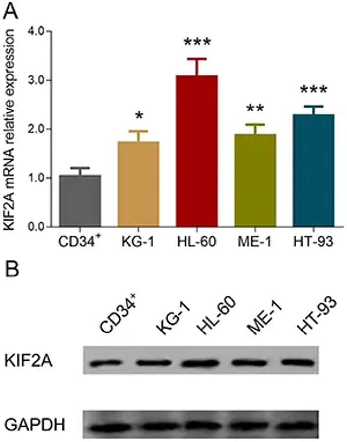
Association of Kinesin Family Member 2A with Increased Disease Risk, Deteriorative Clinical Characteristics, and Shorter Survival Profiles in Acute Myeloid Leukemia
Ding et al. explored KIF2A expression correlation with AML risk, clinical characteristics, and prognosis, and investigated KIF2A knockdown effects on AML cell activities in vitro.
Bone marrow samples from 176 AML patients and 40 healthy donors were analyzed by qPCR. KIF2A was elevated in AML patients and predicted increased disease risk (AUC: 0.793, 95%CI: 0.724-0.826), correlating positively with white blood cells, monosomal karyotype, and high-risk stratification. Kaplan-Meier analysis showed KIF2A negatively correlated with event-free and overall survival. In vitro studies showed that KIF2A mRNA expression was elevated in KG-1, HL-60, ME-1, and HT-93 cell lines compared to CD34+ cells (Fig. 1A). KIF2A protein expression was also increased in KG-1, HL-60, ME-1, and HT-93 cell lines compared to CD34+ cells (Fig. 1B). In conclusion, KIF2A showed potential to be a biomarker and treatment target in AML.
Ask a Question
Write your own review
- You May Also Need

Description: Established in 2007 from the bone marrow mononuclear cells of an 82-year-old Japanese man with diffuse large B-cell lymphoma in the leukemic phase

Description: Established from the bone marrow of a 28-year-old man who developed the terminal leukemic phase of lymphosarcoma in 1976

Description: This cell line was derived from the bone marrow aspirate of a 59 year old male with erythroleukemia that became acute myelogenous leukaemia.The cells form colonies in soft-agar in the presence of ...

Description: Established from the pleural effusion of a 24-year-old woman with recurrent anaplastic large cell lymphoma (ALCL); cells were described to clonally derive from T-lineage lymphoid cells and to be ...

Description: Established from a 37-year-old man at second (refractory/terminal) relapse of Hodgkin lymphoma (nodular sclerosing -> lymphocyte depleted/stage IIISA -> stage IV) after both combined chemo- and ...

Description: Established from the peripheral blood of a 10-year-old Caucasian boy with acute lymphoblastic leukemia (pre B-ALL) at diagnosis in 1993
- Adipose Tissue-Derived Stem Cells
- Human Neurons
- Mouse Probe
- Whole Chromosome Painting Probes
- Hepatic Cells
- Renal Cells
- In Vitro ADME Kits
- Tissue Microarray
- Tissue Blocks
- Tissue Sections
- FFPE Cell Pellet
- Probe
- Centromere Probes
- Telomere Probes
- Satellite Enumeration Probes
- Subtelomere Specific Probes
- Bacterial Probes
- ISH/FISH Probes
- Exosome Isolation Kit
- Human Adult Stem Cells
- Mouse Stem Cells
- iPSCs
- Mouse Embryonic Stem Cells
- iPSC Differentiation Kits
- Mesenchymal Stem Cells
- Immortalized Human Cells
- Immortalized Murine Cells
- Cell Immortalization Kit
- Adipose Cells
- Cardiac Cells
- Dermal Cells
- Epidermal Cells
- Peripheral Blood Mononuclear Cells
- Umbilical Cord Cells
- Monkey Primary Cells
- Mouse Primary Cells
- Breast Tumor Cells
- Colorectal Tumor Cells
- Esophageal Tumor Cells
- Lung Tumor Cells
- Leukemia/Lymphoma/Myeloma Cells
- Ovarian Tumor Cells
- Pancreatic Tumor Cells
- Mouse Tumor Cells