
EFM-192B
Cat.No.: CSC-C0386
Species: Homo sapiens (Human)
Source: Pleural Effusion Metastasis
Morphology: epitheloid adherent cells growing as monolayers
Culture Properties: monolayer
- Specification
- Background
- Scientific Data
- Q & A
- Customer Review
Immunology: cytokeratin +, cytokeratin-7 +, cytokeratin-8 -, cytokeratin-17 +, cytokeratin-18 +, desmin -, endothel -, EpCAM +, GFAP -, neurofilament -, vimentin -
Viruses: EL
EFM-192B cells are a human breast carcinoma cell line originally isolated from pleural effusion of a patient with metastatic breast cancer. These cells are luminal-type breast cancer cells which strongly overexpress and amplify HER2/ERBB2 and are often used as an in vitro tumor model to study HER2-driven cancer biology and response to therapeutics.
Morphologically, EFM-192B cells are epithelial and form adherent monolayers when cultured. At the molecular level, EFM-192B cells generally retain epithelial markers such as cytokeratins and E-cadherin and are usually HER2-positive (i.e. have high levels of HER2 protein expression on the cell surface). As such, these cells are commonly used to test responses to HER2-targeting therapeutics such as monoclonal antibodies or tyrosine kinase inhibitors. EFM-192B cells have also been used to study downstream signaling of HER2 activation such as activation of the PI3K/AKT signaling pathway and MAPK signaling which promote cell proliferation, survival, and drug resistance. These cells have also been used in studies of receptor crosstalk, endocrine resistance, tumor microenvironment, etc. Since they are a cell line, EFM-192B cells can also be used to study mechanisms of oncogenic signaling, gene expression regulation, etc. in a defined genetic background.
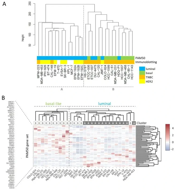
Molecular Subtyping of BC Cell Lines Separate Basal-like from Luminal BC Models
Selecting the most representative breast-cancer (BC) cell line is hampered by incomplete molecular annotation. Pommerenke et al. performed deep profiling (RNA-seq, mutation calling, immunoblotting) of 29 authenticated BC lines to create a comprehensive reference map that links each line to clinical BC subtypes and reveals novel genetic drivers.
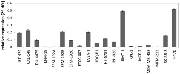
Transcriptome-wide analysis revealed that all basal-like cell lines clustered in group B and were classified as TNBCs, while luminal-expressing cell lines (ESR1⁺, FOXA1⁺) primarily clustered in group A (Fig. 1B). Notably, ERBB2-high cell lines (>200 tpm) including BT-474, EFM-192 variants, IPH-926, JIMT-1, MDA-MB-453, and SK-BR-3 did not form a distinct branch but localized within the luminal arm. All AR⁺ cell lines were confined to the luminal/cluster A group, though JIMT-1 exhibited a mixed phenotype, clustering as HER2⁺ in group B despite luminal features. PAM50 analysis failed to sub-classify luminal cell lines into LumA and LumB, consistent with previous BC cell line studies. We therefore examined miR-99a-5p, a LumA-associated tumor suppressor microRNA. qPCR revealed strong expression in JIMT-1 and T-47D, but absence in EFM-19, EFM-192A, ETCC-007, KPL-1, MCF-7, MDA-MB-453, and MFM-223 (Fig. 2). However, intra-patient heterogeneity was evident: EFM-192B and EFM-192C expressed miR-99a-5p whereas EFM-192A did not, suggesting clonal variation weakens its discriminatory power. Given evidence for subtype admixture in BC, we refrained from LumA/LumB assignment based on transcriptome or miR-99a-5p data alone.
Ask a Question
Write your own review
- You May Also Need

Description: MCF10DCIS.com is a clonal breast cancer cell line derived from a xenograft originating from premalignant MCF10AT cells that were injected into severe combined immune-deficient mice. MCF10AT is ...

Description: Species: human, Caucasian female 69 years old; Tissue: breast; Tumor: adenocarcinoma; Transfected with ER-pBact, human estrogen receptor cDNA, under control of the human beta actin promotor, ...

Description: Human malignant mesothelioma. CA19-9, CA125, and hyaluronic acid producing. Cell growth is slow.

Description: Histopathology: breast cancerNote: NK (natural killer) cell resistant clone

Description: ZR-75-30 was derived from malignant ascites fluid from a 47-year-old premenopausal Black woman with infiltrating ductal carcinoma.

Description: Species: human - female, 74 years old, CaucasianTumorigenecity: yes, in nude miceIsoenzyme: G6PD, B; PGM3, 1; PGM1, 1; ES-D, 1; Me-2, 0; AK-1, 1; GLO-1, 1Histopathology: carcinoma, ductal
- Adipose Tissue-Derived Stem Cells
- Human Neurons
- Mouse Probe
- Whole Chromosome Painting Probes
- Hepatic Cells
- Renal Cells
- In Vitro ADME Kits
- Tissue Microarray
- Tissue Blocks
- Tissue Sections
- FFPE Cell Pellet
- Probe
- Centromere Probes
- Telomere Probes
- Satellite Enumeration Probes
- Subtelomere Specific Probes
- Bacterial Probes
- ISH/FISH Probes
- Exosome Isolation Kit
- Human Adult Stem Cells
- Mouse Stem Cells
- iPSCs
- Mouse Embryonic Stem Cells
- iPSC Differentiation Kits
- Mesenchymal Stem Cells
- Immortalized Human Cells
- Immortalized Murine Cells
- Cell Immortalization Kit
- Adipose Cells
- Cardiac Cells
- Dermal Cells
- Epidermal Cells
- Peripheral Blood Mononuclear Cells
- Umbilical Cord Cells
- Monkey Primary Cells
- Mouse Primary Cells
- Breast Tumor Cells
- Colorectal Tumor Cells
- Esophageal Tumor Cells
- Lung Tumor Cells
- Leukemia/Lymphoma/Myeloma Cells
- Ovarian Tumor Cells
- Pancreatic Tumor Cells
- Mouse Tumor Cells