
DOGKIT
Cat.No.: CSC-C0621
Species: Homo sapiens (Human)
Source: Blood; Peripheral Blood
Morphology: round cells growing singly in suspension
Culture Properties: suspension
- Specification
- Background
- Scientific Data
- Q & A
- Customer Review
Immunology: CD3 -, CD10 +, CD13 -, CD19 +, CD20 +, CD34 -, CD37 +, CD38 +, cyCD79a +, CD80 -, CD138 +, sm/cyIgG -, sm/cyIgM -, sm/cykappa -, sm/cylambda -
Viruses: PCR: EBV -, HBV -, HCV -, HIV -
DOGKIT was derived from two patients with chemoresistant B-cell lymphoma, and is an aggressive lymphoma cell line able to grow in vitro. It has been used as an in vitro model system for B-cell lymphomas that mimic disease failure and relapse. DOGKIT cells are cultured in suspension, and demonstrate a morphology that is indicative of lymphoid cells. Immunophenotyping of DOGKIT cells reveals expression of markers indicative of B-cell origin, demonstrating derivation from a B-cell lymphoma. Behaviorally, DOGKIT cells demonstrate continued proliferation, as well as prolonged survival when exposed to cytotoxic stress. In general, these properties recapitulate the resistant phenotype exhibited by the tumor from which DOGKIT was derived.
Gene expression profiling of DOGKIT cells revealed alterations in pathways associated with cell cycle progression, evasion of apoptosis, and DNA damage repair. These changes are often linked with resistance to chemotherapy in B-cell lymphomas, and lead to generalized insensitivity to cytotoxic chemotherapy drugs. Due to these properties, DOGKIT cells have been utilized in studies looking at drug resistance caused by changes in apoptotic signaling cascades, changes in drug efflux pumps, and other survival pathways.
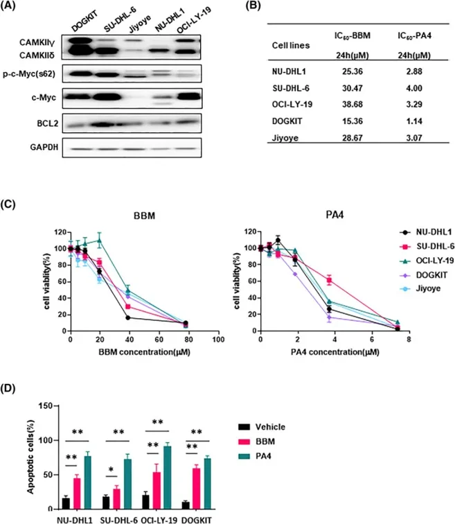
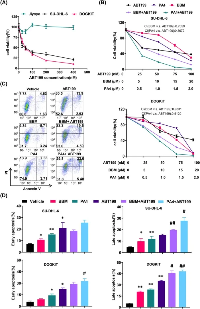
BBM or PA4 Effectively Inhibited DHL Cell Growth and Induced Cell Apoptosis
Double-hit lymphoma (DHL) with concurrent MYC and BCL2 translocations carries a dismal prognosis. Liu's team investigated whether CaMKIIδ/γ, which are highly expressed in DHL, sustain c-Myc and BCL2 oncoproteins and thus represent actionable therapeutic targets.
Expression analysis revealed that four DHL cell lines exhibited higher levels of CAMKIIδ/γ, c-Myc, and BCL2 compared to Burkitt's lymphoma Jiyoye cells (Fig. 1A). Both BBM and PA4 inhibited DHL cell growth dose-dependently, with PA4 showing markedly lower IC50 values, indicating greater potency (Fig. 1B, C). PA4 also induced substantially higher apoptosis rates (72.67%-91.57% Annexin V-positive) compared to BBM (30%-50%) after 24 h treatment (Figure 2D). The BCL2 inhibitor ABT199 showed growth inhibition in DOGKIT and SU-DHL-6 (Fig. 2A). Drug combination studies revealed distinct synergistic profiles: BBM + ABT199 showed limited synergy (CI50 = 0.7978 in SU-DHL-6), whereas PA4 + ABT199 demonstrated strong synergy (CI50 = 0.3672 in SU-DHL-6; CI50 = 0.5120 in DOGKIT) (Fig. 2B). Neither combination produced clear synergistic effects on apoptosis, though modest increases in early apoptotic cells were observed with BBM + ABT199 in DOGKIT and with both combinations in SU-DHL-6 (Fig. 2C, D).
Ask a Question
Write your own review
- You May Also Need

Description: Established in 2007 from the bone marrow mononuclear cells of an 82-year-old Japanese man with diffuse large B-cell lymphoma in the leukemic phase

Description: Established from the bone marrow of a 28-year-old man who developed the terminal leukemic phase of lymphosarcoma in 1976

Description: This cell line was derived from the bone marrow aspirate of a 59 year old male with erythroleukemia that became acute myelogenous leukaemia.The cells form colonies in soft-agar in the presence of ...

Description: Established from the pleural effusion of a 24-year-old woman with recurrent anaplastic large cell lymphoma (ALCL); cells were described to clonally derive from T-lineage lymphoid cells and to be ...

Description: Established from a 37-year-old man at second (refractory/terminal) relapse of Hodgkin lymphoma (nodular sclerosing -> lymphocyte depleted/stage IIISA -> stage IV) after both combined chemo- and ...

Description: Established from the peripheral blood of a 10-year-old Caucasian boy with acute lymphoblastic leukemia (pre B-ALL) at diagnosis in 1993
- Adipose Tissue-Derived Stem Cells
- Human Neurons
- Mouse Probe
- Whole Chromosome Painting Probes
- Hepatic Cells
- Renal Cells
- In Vitro ADME Kits
- Tissue Microarray
- Tissue Blocks
- Tissue Sections
- FFPE Cell Pellet
- Probe
- Centromere Probes
- Telomere Probes
- Satellite Enumeration Probes
- Subtelomere Specific Probes
- Bacterial Probes
- ISH/FISH Probes
- Exosome Isolation Kit
- Human Adult Stem Cells
- Mouse Stem Cells
- iPSCs
- Mouse Embryonic Stem Cells
- iPSC Differentiation Kits
- Mesenchymal Stem Cells
- Immortalized Human Cells
- Immortalized Murine Cells
- Cell Immortalization Kit
- Adipose Cells
- Cardiac Cells
- Dermal Cells
- Epidermal Cells
- Peripheral Blood Mononuclear Cells
- Umbilical Cord Cells
- Monkey Primary Cells
- Mouse Primary Cells
- Breast Tumor Cells
- Colorectal Tumor Cells
- Esophageal Tumor Cells
- Lung Tumor Cells
- Leukemia/Lymphoma/Myeloma Cells
- Ovarian Tumor Cells
- Pancreatic Tumor Cells
- Mouse Tumor Cells