
ARH-77
Cat.No.: CSC-C0521
Species: Homo sapiens (Human)
Source: Blood; Peripheral Blood
Morphology: round single cells or clustered growing in suspension, some cells loosely adherent
Culture Properties: suspension
- Specification
- Background
- Scientific Data
- Q & A
- Customer Review
D13S317: 11,13
D7S820: 7,12
D16S539: 9,13
vWA: 17
THO1: 8,9.3
Amelogenin: X
TPOX: 8
CSF1PO: 6,10
ARH-77 is a human B-lymphoblastoid cell line that was first isolated from peripheral blood taken from a 33-year-old woman with IgG plasma cell leukemia. It was originally suggested to be a myeloma/plasma cell leukemia cell line. However, further characterization has shown that ARH-77 is an Epstein-Barr virus (EBV)-transformed B lymphoblastoid line, and not a true malignant plasma cell line. ARH-77 cells are grown in suspension as either round cells or cell clusters that may or may not loosely adhere to each other.
Phenotypically, ARH-77 expresses CD11a, CD19, CD20, CD28, CD49e, and does not express CD38. It produces IgG1 with kappa light chain. It is EBV-positive and contains latent viral nuclear and capsid antigens, but does not lytically replicate. In initial studies, ARH-77 was shown to strongly proliferate in immunoglobulin-secreting cell numbers when stimulated by either lymphokines or phorbol myristate acetate (PMA). Isotype switching does not occur. Therefore, ARH-77 has been used as an in vitro model to study B-cell activation and mechanisms of IgG secretion. ARH-77, or its engineered derivatives, are also commonly used in immuno-oncology; they are the host cells for many cytotoxicity assays (ADCC, CDC, ADCP) used in the study of antibody-based and cell-based therapeutics. Fast-growing mutants of ARH-77 (HMy2) have also been incorporated in hybridoma systems for monoclonal antibody production.
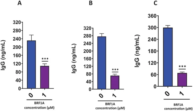
BRF1A Inhibits Excessive IgG Production in ARH77 Cells without Cytotoxicity
Myeloma cells relentlessly secrete IgE/IgG and rely on NF-κB, c-Myc, and telomerase for survival. Musa et al. evaluated BRF1A, a cannabidiol-enriched extract, for its ability to curb malignant Ig secretion and growth of U266/ARH-77 myeloma cells.
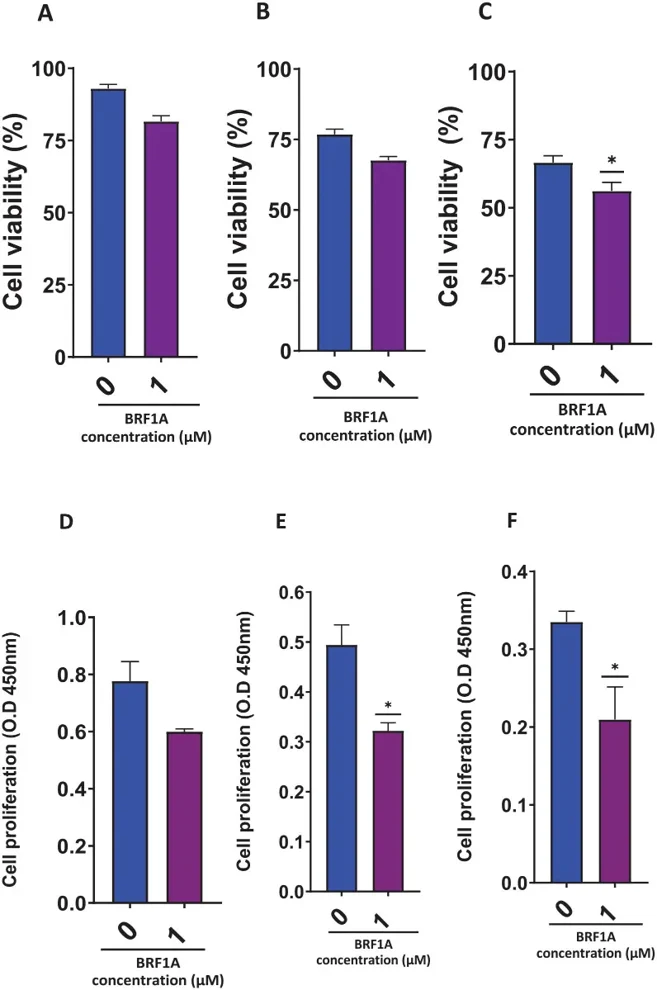
Musa et al. found that BRF1A suppressed IgE-producing myeloma function, therefore they wanted to see if it had a similar effect on IgG-producing myeloma ARH-77 cells. They cultured ARH-77 cells with 1.0 µM BRF1A for 1, 3, and 5 days. Compared to untreated cultures, IgG production significantly decreased on days 1, 3, and 5 (Fig. 1A-C). Within 24 hours, the IgG concentration in BRF1A-cultured cells was 79 to 144 ng/mL, compared to 185 to 299 ng/mL in untreated cells. The IgG concentration continued to decrease on days 3 and 5. On day 3, the treatment group had IgG concentrations of 46 to 99 ng/mL, while the untreated group had 244 to 316 ng/mL. On day 5, the treatment group had 44 to 94 ng/mL, and the untreated group had 278 to 329 ng/mL. No significant cytotoxicity was observed on days 1 and 3 (Fig. 2A-C). Additionally, CCK-8 assays showed that BRF1A had a similar effect on IgG-producing myeloma cells as on IgE-producing cells (Fig. 2D-F). Thus, BRF1A can suppress both IgE- and IgG-producing myeloma cells, highlighting its potential as a therapeutic agent.
Ask a Question
Write your own review
- You May Also Need

Description: Established in 2007 from the bone marrow mononuclear cells of an 82-year-old Japanese man with diffuse large B-cell lymphoma in the leukemic phase

Description: Established from the bone marrow of a 28-year-old man who developed the terminal leukemic phase of lymphosarcoma in 1976

Description: This cell line was derived from the bone marrow aspirate of a 59 year old male with erythroleukemia that became acute myelogenous leukaemia.The cells form colonies in soft-agar in the presence of ...

Description: Established from the pleural effusion of a 24-year-old woman with recurrent anaplastic large cell lymphoma (ALCL); cells were described to clonally derive from T-lineage lymphoid cells and to be ...

Description: Established from a 37-year-old man at second (refractory/terminal) relapse of Hodgkin lymphoma (nodular sclerosing -> lymphocyte depleted/stage IIISA -> stage IV) after both combined chemo- and ...

Description: Established from the peripheral blood of a 10-year-old Caucasian boy with acute lymphoblastic leukemia (pre B-ALL) at diagnosis in 1993
- Adipose Tissue-Derived Stem Cells
- Human Neurons
- Mouse Probe
- Whole Chromosome Painting Probes
- Hepatic Cells
- Renal Cells
- In Vitro ADME Kits
- Tissue Microarray
- Tissue Blocks
- Tissue Sections
- FFPE Cell Pellet
- Probe
- Centromere Probes
- Telomere Probes
- Satellite Enumeration Probes
- Subtelomere Specific Probes
- Bacterial Probes
- ISH/FISH Probes
- Exosome Isolation Kit
- Human Adult Stem Cells
- Mouse Stem Cells
- iPSCs
- Mouse Embryonic Stem Cells
- iPSC Differentiation Kits
- Mesenchymal Stem Cells
- Immortalized Human Cells
- Immortalized Murine Cells
- Cell Immortalization Kit
- Adipose Cells
- Cardiac Cells
- Dermal Cells
- Epidermal Cells
- Peripheral Blood Mononuclear Cells
- Umbilical Cord Cells
- Monkey Primary Cells
- Mouse Primary Cells
- Breast Tumor Cells
- Colorectal Tumor Cells
- Esophageal Tumor Cells
- Lung Tumor Cells
- Leukemia/Lymphoma/Myeloma Cells
- Ovarian Tumor Cells
- Pancreatic Tumor Cells
- Mouse Tumor Cells