FISH Protocol for Interphase Chromosomes
GUIDELINE
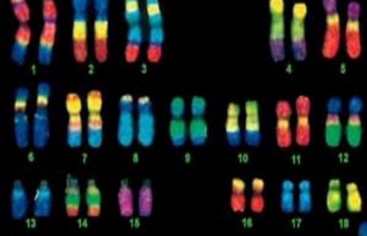
Interphase fluorescence in situ hybridization (I-FISH) is a useful technique for detecting chromosomal numerical abnormalities in tumors and is gaining acceptance as a tool in cytogenetics and clinical diagnoses.
METHODS
- Remove Coplin jar(s) containing slides from 4°C allow them to warm to room temperature.
- Incubate slides in 2×SSCT + 50% (v/v) formamide for 2.5 min at 92°C in a prewarmed Coplin jar.
- Transfer slides to a Coplin jar containing 2×SSCT + 50% formamide at 60°C and incubate for 20 min.
- Remove the slides from the 60°C Coplin jar and allow them to cool to room temperature.
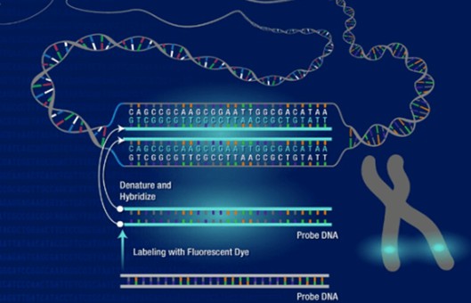
- Add 25 µl of a hybridization cocktail composed of 2×SSCT, 50% formamide, 10% (w/v) dextran sulfate, 10 µg RNase A, and 10-20 pmole of Oligopaints probe to a 22x22 #1.5 coverslip.
- Invert slides onto the cocktail-containing coverslips such that the area containing the cells is covered. Seal with rubber cement.
- Allow the rubber cement to air-dry for 5 min at room temperature.
- Denature for 2.5 min at 92°C by placing slides on top of a water-immersed heat block inside a water bath.
- Transfer slides to a humidified chamber and hybridize overnight at 37°C or 42°C.
- The next day, remove the coverslip carefully and wash the slides in a pre-warmed Coplin jar containing 2×SSCT at 60°C for 15 min.
- Transfer slides to a Coplin jar containing 2×SSCT at room temperature and incubate for 10 min.
- Transfer slides to a Coplin jar containing 0.2X SSC at room temperature and incubate for 10 min.
- Remove slides from the Coplin jar and gently tap them dry on paper towels – take care to just tap the thin edge of the slide and to never allow the surface containing the cells to directly contact the paper towels.
- Add 15 µl of mounting media such as SlowFade Gold + DAPI to a 22x30 #1.5 coverslip.
- Invert slides onto mounting media-containing coverslips such that the area containing the cells is centered beneath the coverslip.
- Seal each slide using nail polish.
- Allow at least 30 min for the nail polish to dry before imaging the slides to ensure no nail polish gets on the microscope objectives.
- In our hands, slides remain quite stable (e.g. for several months) when stored at 4°C and protected from light.
Creative Bioarray Relevant Recommendations
- Compared with interim analysis, the main advantage of interphase cytogenetics is that it does not require cell culture, making the procedure directly applicable to cell materials and allowing more cells to be analyzed more quickly. Creative Bioarray provides Interphase FISH Services that can be used to analyze a variety of cell nuclei. Our IFISH service uses advanced peptide nucleic acid (PNA) probes for better hybridization results.
NOTES
- If using 2 hybridization areas per slide, separate probe areas with a streak of nail polish or pap pen to keep probe solutions from mixing.
- Do not let the mounting solution come to room temperature before removing the aliquot; keep it at 4°C until ready to use. DAPI staining intensity will correspond inversely to the degree of chromosomal denaturation.