
V-79
Cat.No.: CSC-C2756
Species: Cricetulus griseus (Chinese hamster)
Source: Lung
Morphology: fibroblast
Culture Properties: adherent
- Specification
- Background
- Scientific Data
- Q & A
- Customer Review
V-79 cells (also known as V79 or V79-4 cells) are a fibroblast-like Chinese hamster cell line developed from lung fibroblasts of the Chinese hamster Cricetulus griseus. Since their development, V-79 cells have been one of the most commonly used mammalian cell lines for genetic toxicology and radiobiology assays. The cell line has been shown to have a stable karyotype and proliferate quickly with high plating efficiency.
In culture, V-79 cells are an adherent monolayer cell line with rapid doubling time and high clonogenic survival. These cells are used for mutation assays, DNA damage assays, and cell survival assays. Because of their low endogenous metabolic enzyme activity, induction of mutation and DNA damage by test compounds can be easily evaluated either directly or after incubation with an exogenous metabolic activation system like liver S9 fractions. For these reasons, V-79 cells have been commonly used for HPRT gene mutation assays, chromosomal aberration assays, and micronucleus assays.
V-79 cells have been extensively used in research of radiation biology studies along with oxidative stress induction and DNA repair mechanisms. Because of their consistent responses to chemicals and radiation, easy culturability, and stability over extended periods of time, V-79 cells continue to be a preferred in vitro model system for testing cytotoxicity and mutagenicity as well as determining cellular responses to environmental chemicals and therapeutics.
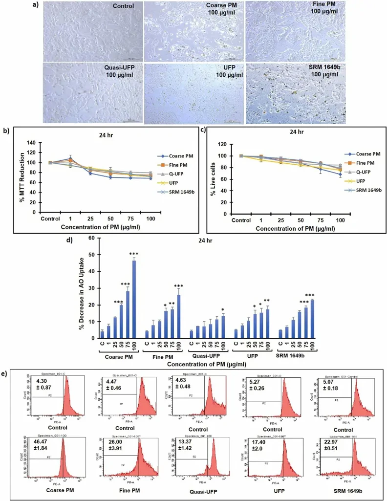
Cytotoxicity and Cell Viability Induced by PM in V-79 Cells
Particulate Matter (PM) in ambient air is a major environmental and health concern. Dubey et al. aimed to comprehensively characterize atmospheric PM fractions by size, composition, and morphology, and assess their oxidative potential, genotoxicity, and mutagenicity near a busy roadside in Lucknow, India.
After being exposed to PM for 24 hours, adherent and settled particulate on the surface of V-79 cells had no visible effects on cellular morphology (Fig. 1a). Cells that were exposed to coarse PM became irregular in shape, shrunken, and lifted off the surface in some cases. PM demonstrated a significant concentration-dependent cytotoxic effect (1-100 µg/ml) after 24 hours of exposure, according to cytotoxicity determined by MTT assay. Coarse PM produced the greatest cytotoxicity when compared to other PM fractions and SRM-1649b (Fig. 1b). There was a significant increase in Trypan blue uptake by cells exposed to PM concentrations of 50, 75, and 100 µg/ml after 24 hours of exposure when compared to the control, demonstrating lower cell viability. Coarse PM-exposed cells displayed the greatest loss in cell viability (Fig. 1c). When compared to untreated control cells, acridine orange staining demonstrated concentration-dependent loss in lysosome membrane integrity. Coarse fraction-exposed cells displayed the greatest loss in membrane integrity (Fig. 1d, e).
Ask a Question
Write your own review
- You May Also Need

Description: Established from the BHP-induced pancreatic carcinoma of a Syrian golden hamster (Mesocricetus auratus)(GN strain) in 1983


Description: Established from the lung of a Syrian golden hamster fetus on day 15 after gestation in 1981; primary culture cells were treated with hydroxy-proline; cells were described as premature epithelial ...

Description: Species: hamster, Chinese female adult; Tissue: ovary; Transfected with the SSR1 somatostatin receptor; Original line: CHO-K1
- Adipose Tissue-Derived Stem Cells
- Human Neurons
- Mouse Probe
- Whole Chromosome Painting Probes
- Hepatic Cells
- Renal Cells
- In Vitro ADME Kits
- Tissue Microarray
- Tissue Blocks
- Tissue Sections
- FFPE Cell Pellet
- Probe
- Centromere Probes
- Telomere Probes
- Satellite Enumeration Probes
- Subtelomere Specific Probes
- Bacterial Probes
- ISH/FISH Probes
- Exosome Isolation Kit
- Human Adult Stem Cells
- Mouse Stem Cells
- iPSCs
- Mouse Embryonic Stem Cells
- iPSC Differentiation Kits
- Mesenchymal Stem Cells
- Immortalized Human Cells
- Immortalized Murine Cells
- Cell Immortalization Kit
- Adipose Cells
- Cardiac Cells
- Dermal Cells
- Epidermal Cells
- Peripheral Blood Mononuclear Cells
- Umbilical Cord Cells
- Monkey Primary Cells
- Mouse Primary Cells
- Breast Tumor Cells
- Colorectal Tumor Cells
- Esophageal Tumor Cells
- Lung Tumor Cells
- Leukemia/Lymphoma/Myeloma Cells
- Ovarian Tumor Cells
- Pancreatic Tumor Cells
- Mouse Tumor Cells