
Human Mesenchymal Stem Cells from Umbilical Cord Matrix (hMSC-UC)
- Specification
- Background
- Scientific Data
- Q & A
- Customer Review
Human Mesenchymal Stem Cells from Umbilical Cord Matrix (hMSC-UC), also known as Wharton's jelly-derived mesenchymal stem cells (UC-MSCs), are multipotent stromal cells that have been isolated from the gelatinous connective tissue of the human umbilical cord. These cells have fibroblast-like form, high self-renewal capacity and multilineage differentiation potential, allowing them to differentiate into osteogenic, chondrogenic, adipogenic and other mesenchymal lineages under adequate culture conditions.
hMSC-UCs express normal mesenchymal markers such as CD73, CD90, and CD105, and do not express haematopoietic markers such as CD34 and CD45. Compared with MSCs obtained from adult tissues, umbilical cord-derived MSCs have a stronger proliferative potential, reduced immunogenicity and a more basic developmental state, making them particularly appealing for regenerative medicine and cell therapy applications.
Functionally, hMSC-UCs exert significant immunomodulatory and paracrine effects (e.g., release of cytokines, growth factors, and extracellular vesicles) that control inflammation and promote tissue repair and angiogenesis. These features have stimulated their extensive examination in pre-clinical and clinical investigations for tissue regeneration, immunological disorders, neurodegenerative diseases and inflammatory conditions.
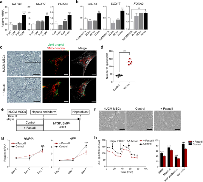
Fasudil Induces Early Hepatic Endoderm Differentiation of hUCM-MSCs Inhibition of Further Hepatoblast Maturation.
Human umbilical cord matrix mesenchymal stem cells (hUCM-MSCs) are a promising source for tissue regeneration, however efficient differentiation techniques are still limited. This study examined the ROCK inhibitor fasudil for hepatic differentiation. Screening indicated that 10 µM fasudil for 72 hours was optimum and significantly increased endoderm markers (GATA4, SOX17, FOXA2) compared to controls (Fig. 1a, b). However, fasudil was more effective at day 3 (Fig. S1a and b) than other ROCK inhibitors (Y-27632, ripasudil), which also elevated these genes.
Tomographic studies showed that fasudil-treated cells were more ovoid in form with changed mitochondrial morphology and a considerable increase in lipid droplet (LD) densities (Fig. 1c, d). However, whereas endoderm gene expression was boosted, subsequent hepatoblast differentiation was inhibited. On day 7, the hepatoblast marker AFP was dramatically decreased in the fasudil group, whereas HNF4A was unaffected (Fig. 1e-g). This inhibition was associated with reduced mitochondrial activity at day 3 (Fig. 1h), possibly caused by an excessive accumulation of LDs interfering with energy production. Fasudil was superior to other inhibitors in preserving ATP levels (Fig. S1c, d), although its creation of LDs ultimately impeded differentiation at later stages. These results suggest that fasudil stimulates transcriptional activation of early endoderm but disturbs organelle homeostasis, preventing their further development into hepatoblasts.
Ask a Question
Write your own review
- You May Also Need
- Adipose Tissue-Derived Stem Cells
- Human Neurons
- Mouse Probe
- Whole Chromosome Painting Probes
- Hepatic Cells
- Renal Cells
- In Vitro ADME Kits
- Tissue Microarray
- Tissue Blocks
- Tissue Sections
- FFPE Cell Pellet
- Probe
- Centromere Probes
- Telomere Probes
- Satellite Enumeration Probes
- Subtelomere Specific Probes
- Bacterial Probes
- ISH/FISH Probes
- Exosome Isolation Kit
- Human Adult Stem Cells
- Mouse Stem Cells
- iPSCs
- Mouse Embryonic Stem Cells
- iPSC Differentiation Kits
- Mesenchymal Stem Cells
- Immortalized Human Cells
- Immortalized Murine Cells
- Cell Immortalization Kit
- Adipose Cells
- Cardiac Cells
- Dermal Cells
- Epidermal Cells
- Peripheral Blood Mononuclear Cells
- Umbilical Cord Cells
- Monkey Primary Cells
- Mouse Primary Cells
- Breast Tumor Cells
- Colorectal Tumor Cells
- Esophageal Tumor Cells
- Lung Tumor Cells
- Leukemia/Lymphoma/Myeloma Cells
- Ovarian Tumor Cells
- Pancreatic Tumor Cells
- Mouse Tumor Cells