
H-209
Cat.No.: CSC-C0510
Species: Homo sapiens (Human)
Source: Bone Marrow Metastasis
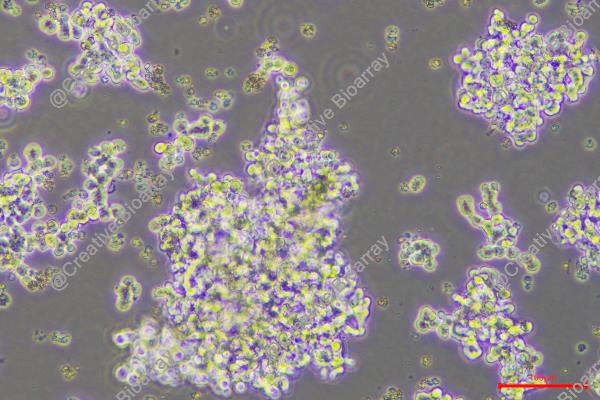
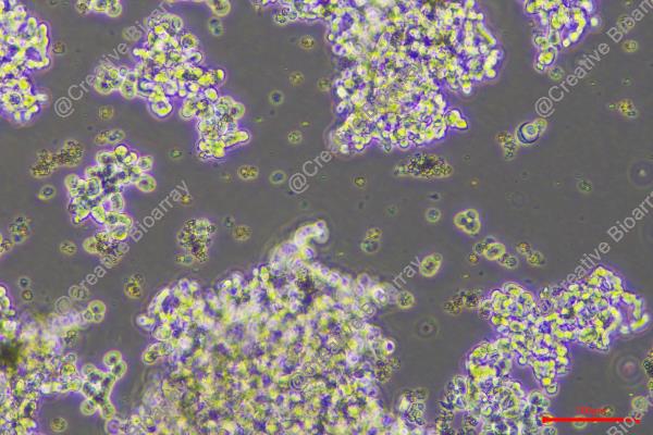
Morphology: spherical-to-ovoid cells growing singly in large clumps
- Specification
- Background
- Scientific Data
- Q & A
- Customer Review
Immunology: cytokeratin +, cytokeratin-7 -, cytokeratin-8 +, cytokeratin-17 -, cytokeratin-18 +, cytokeratin-19 +, desmin -, endothel -, EpCAM +, GFAP -, neurofilament -, vimentin -
Viruses: PCR
The H209 cell line, also known as NCI-H209, is a classical human Small Cell Lung Cancer (SCLC) cell line developed by Gazdar and coworkers in 1979. It was obtained from the site of metastasis of a 55-year-old white male in the bone marrow before any treatment. The line is genetically hyperdiploid with a median chromosomal number of 49 and has homozygous mutations in RB1(p.Cys706Phe) and TP53(c.673-2A>T) gene.
In H209 cells display an unusual growth behavior in culture, growing as tight, spherical aggregates in suspension instead of attaching to the flask surface. Only cells within these aggregates remain alive, but the culture media generally contains a considerable quantity of cellular debris, a normal characteristic of this line. H209 is a neuroendocrine tumor model that expresses high levels of typical SCLC indicators such neuron-specific enolase, creatine kinase-BB, L-DOPA decarboxylase, and bombesin-like immunoreactivity.
It is normally cultured in a serum-supplemented rich basal media under conventional conditions (37°C, 5% CO2). The H209 line is widely used in oncology research for the study of SCLC pathology, neuroendocrine differentiation, drug screening and tumor biology due to its unique biological characteristics.
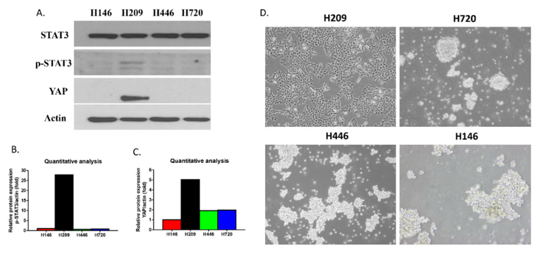
STAT3 and YAP Expression in Small Cell Lung Cancer Lines
Small cell lung cancer (SCLC) is an aggressive malignancy with poor prognosis, yet the interplay between key oncogenic pathways remains unclear. While STAT3 and YAP are established drivers of tumor progression, their potential interaction in SCLC pathogenesis is undefined.
Western blot analysis revealed that among four SCLC cell lines, H209 cells uniquely exhibited concurrent high expression of both phosphorylated STAT3 (p-STAT3) and YAP protein compared to H146, H446, and H720 cells (Figure 1A-C). This molecular profile correlated with distinct phenotypic behavior: H209 cells displayed an adherent growth pattern, whereas the other cell lines with low p-STAT3/YAP expression exhibited a characteristic floating growth pattern (Figure 1D). These data suggest a potential link between STAT3/YAP signaling and the adhesive phenotype in SCLC.
Ask a Question
Write your own review
- You May Also Need

Description: Lung small cell carcinoma producing insulin-like growth factor II. Cell growth is slow.

Description: Lung Cancer-1/squamous. Parent cell line of LC-1/sq-SF, the same patient as LC-F. Cell growth is slow.

Description: Species: human - male, 47 years old, CaucasianTumorigenecity: does not produce tumorsIsoenzyme: Me-2, 1-2; PGM3, 1-2; PGM1, 1; ES D, 2; AK1, 1; GLO-1, 1-2; G6PD, BHistopathology: carcinoma, ...


Description: Derived from the lung of a 56-year-old black man with small cell lung carcinoma prior to treatment in 1988
- Adipose Tissue-Derived Stem Cells
- Human Neurons
- Mouse Probe
- Whole Chromosome Painting Probes
- Hepatic Cells
- Renal Cells
- In Vitro ADME Kits
- Tissue Microarray
- Tissue Blocks
- Tissue Sections
- FFPE Cell Pellet
- Probe
- Centromere Probes
- Telomere Probes
- Satellite Enumeration Probes
- Subtelomere Specific Probes
- Bacterial Probes
- ISH/FISH Probes
- Exosome Isolation Kit
- Human Adult Stem Cells
- Mouse Stem Cells
- iPSCs
- Mouse Embryonic Stem Cells
- iPSC Differentiation Kits
- Mesenchymal Stem Cells
- Immortalized Human Cells
- Immortalized Murine Cells
- Cell Immortalization Kit
- Adipose Cells
- Cardiac Cells
- Dermal Cells
- Epidermal Cells
- Peripheral Blood Mononuclear Cells
- Umbilical Cord Cells
- Monkey Primary Cells
- Mouse Primary Cells
- Breast Tumor Cells
- Colorectal Tumor Cells
- Esophageal Tumor Cells
- Lung Tumor Cells
- Leukemia/Lymphoma/Myeloma Cells
- Ovarian Tumor Cells
- Pancreatic Tumor Cells
- Mouse Tumor Cells