Mouse Conjunctival Fibroblasts
Cat.No.: CSC-C5408S
Species: Mouse
Source: Conjunctiva; Eye
Cell Type: Fibroblast
- Specification
- Background
- Scientific Data
- Q & A
- Customer Review
Mouse conjunctival fibroblasts from Creative Bioarray are isolated from the mouse eye tissue. The method we use to isolate mouse conjunctival fibroblasts was developed based on a combination of established and our proprietary methods. The mouse conjunctival fibroblasts are characterized by immunofluorescence with antibodies specific to desmin or vimentin. Each vial contains 0.5x10^6 cells per ml and is delivered frozen.
The conjunctival fibroblasts of mouse are extracted from the conjunctival tissue that envelops the eye's outer surface particularly in the bulbar conjunctiva where the cornea meets the sclera. These spindle or star-shaped cells release extracellular matrix elements which help preserve eye tissue stability and facilitate repair processes. Their cytoplasm shows no lipid granules when viewed under high magnification which differentiates them from skin fibroblasts. These cells grow quickly when culture conditions are applied and assemble into multilayered structures. They serve as crucial tools in ocular inflammation research through conjunctival inflammation simulation when exposed to inflammatory factors IL-4 and IL-13 in culture. Their pluripotent nature allows scientists to transform these cells into pluripotent stem cells which are used for regenerative medicine and tissue repair.
Promotion of Conjunctival Fibroblast-Mediated Collagen Gel Contraction by Mast Cells through Up-Regulation of Matrix Metalloproteinase Release and Activation
Conjunctival fibroblasts manage ECM homeostasis, critically involved in wound healing and fibrosis, primarily regulated by matrix metalloproteinases (MMPs). Mast cells, prevalent in conjunctival tissues, contribute to allergic reactions and scarring. Utilizing a 3D collagen gel culture system, Kiahimoto et al. investigated fibroblast-mast cell interactions' impact on conjunctival fibroblast contractility and ECM remodeling. Assessments include cell viability, collagen contraction, and MMP activity.
Various numbers of mast cells, alone or with conjunctival fibroblasts, were incubated, and MMP levels were tested using gelatin zymography (Fig. 1). Mast cells alone did not produce MMP-2 or MMP-9, but conjunctival fibroblasts did. Mast cells increased pro-MMP-9 and MMP-2 levels in fibroblasts, linked to the number of mast cells. This suggests mast cells aid pro-MMP-9 release by fibroblasts and help convert pro-MMP-2 to active MMP-2. They explored MMP inhibition on dilation by mast cells. Collagen gels with mast cells or fibroblasts were treated with MMP inhibitor batimastat. Batimastat did not affect fibroblast activity alone but reduced contraction in co-cultures with mast cells (Fig. 2). This implies that mast cell enhancement of contraction depends on MMPs.
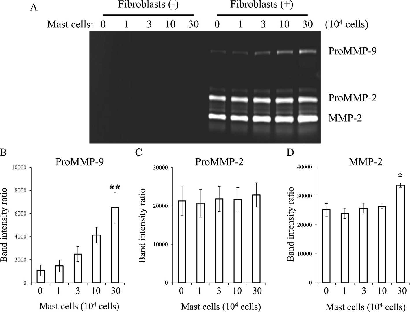 Fig. 1. Effect of mast cells on MMP expression by conjunctival fibroblasts (Kishimoto T, Ishida W, et al., 2022).
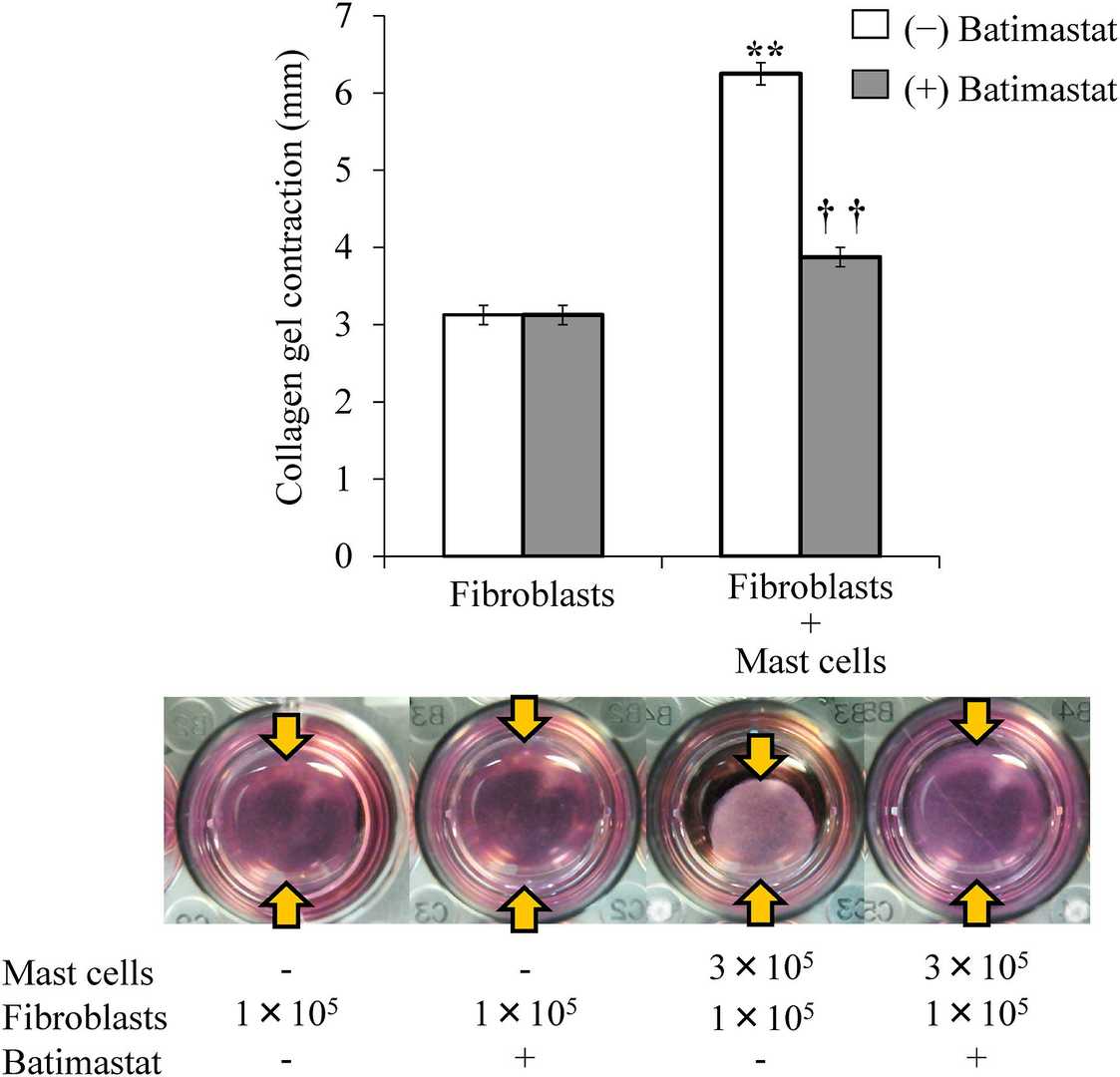 Fig. 2. Effect of batimastat on collagen gel contraction in co-cultures of conjunctival fibroblasts and mast cells (Kishimoto T, Ishida W, et al., 2022).
Increasing the Single siRNA Layer Load in LbL Nanoparticles Prolonged Gene-Silencing Activity in Primary Conjunctival Fibroblasts
The therapeutic use of RNA interference (RNAi) for gene silencing has emerged as a promising treatment for various diseases due to its specificity and low cost. Despite intense research, gene silencing therapies are limited by inefficient delivery systems that degrade or clear siRNA too rapidly. Tan et al. introduced a novel nanocarrier with a hydroxyapatite core and layers of siRNA and cationic peptide to effectively deliver and sustain siRNA targeting SPARC protein. The delivery system was evaluated using FRET studies and in vitro knockdown assays in fibroblasts.
To create siRNA films with high loading and minimal toxicity, hydroxyapatite (HA) was used as the nanoparticle core with alternating layers of ARG, a polycation, and negatively charged siRNA, forming siRNA-LbL films. This method proved effective for cell entry, maintaining viability, and low cytotoxicity. The amount of siRNA in the nanoparticles influences the duration of gene silencing. Three-layered LbL nanoparticles with increased siRNA loads were tested to silence SPARC in mouse conjunctival cells. Nanoparticles with 1.2 pmol SPARC siRNA per μg HA achieved significant mRNA reduction after 7 days (Fig. 3a) and maintained it for 14 days (Fig. 3b). However, the effect lessened over time due to gradual siRNA release. Type I collagen expression remained unchanged (Fig. 3c and d), signifying the need for adjusting dosing and release for sustained silencing.
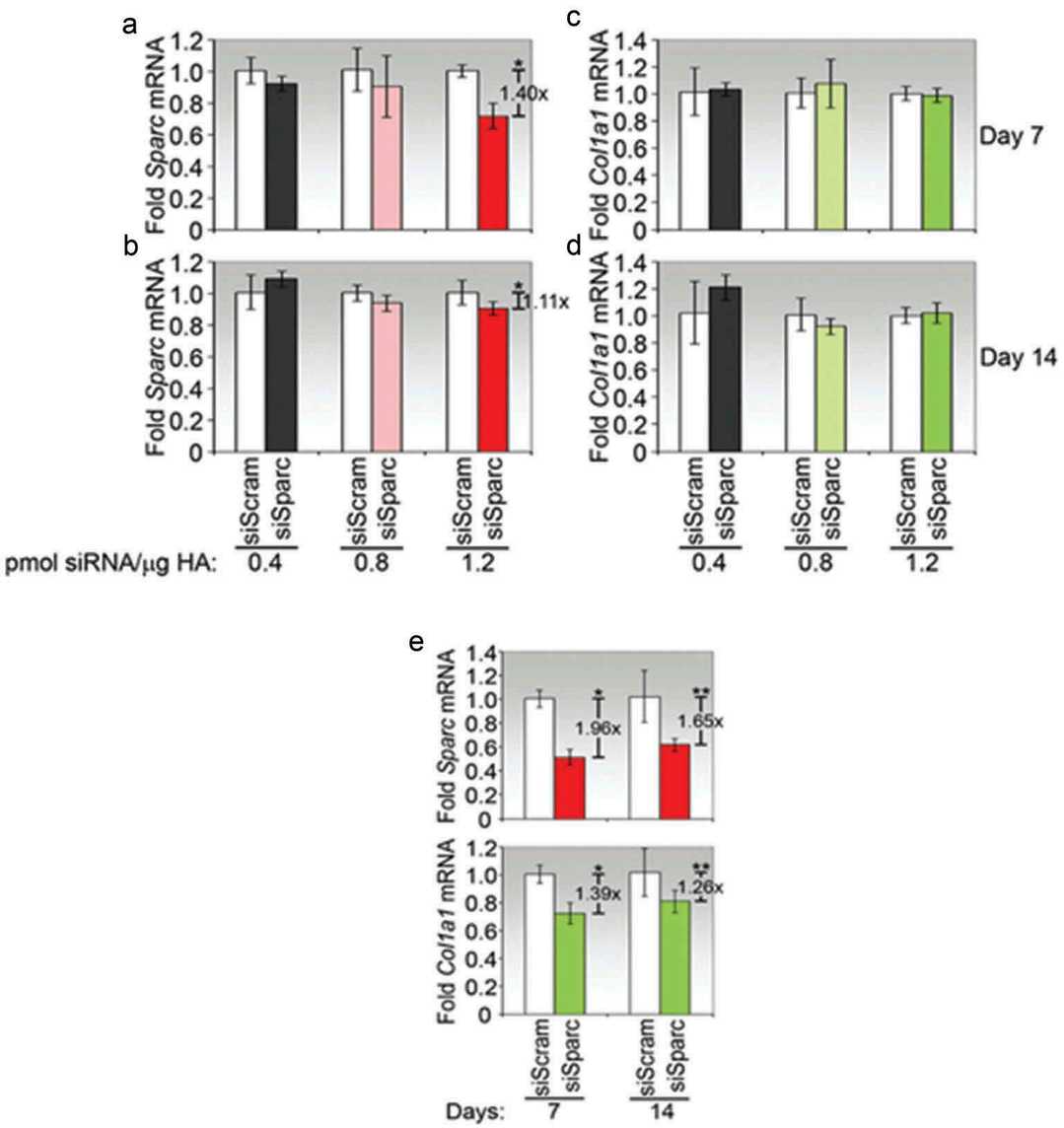 Fig. 3. Ncreasing the single siRNA layer load in LbL nanoparticles prolonged gene-silencing activity in primary conjunctival fibroblasts (Tan Y F, Lee Y S, et al., 2018).
Ask a Question
Write your own review
- You May Also Need


Description: C57BL/6-GFP Mouse Skeletal Muscle Microvascular Endothelial Cells from Creative Bioarray are isolated from C57BL/6-Tg (CAG-EGFP) 1Osb/J mouse skeletal muscle tissue of pathogen-free laboratory mice. ...
Description: eNOS KO Mouse Stomach Epithelial Cells from Creative Bioarray are isolated from stomach tissue of pathogen-free laboratory mice. eNOS KO Mouse Stomach Epithelial Cells are grown in a T25 tissue ...

Description: eNOS KO Mouse Liver Fibroblasts from Creative Bioarray are isolated from liver tissue of pathogen-free laboratory mice. eNOS KO Mouse Liver Fibroblasts are grown in T75 tissue culture flasks ...
Description: C57BL/6-GFP Mouse Corneal Epithelial Cells from Creative Bioarray are isolated from C57BL/6-GFP-Tg(CAG-EGFP)1Osb/J mouse corneal tissue of pathogen-free laboratory mice. C57BL/6-GFP Mouse Corneal ...

Description: BALB/c Mouse Retinal Microvascular Endothelial Cells from Creative Bioarray are isolated from retinal tissue of pathogen-free laboratory mice. BALB/c Mouse Retinal Microvascular Endothelial Cells are ...