Featured Products
Our Promise to You
Guaranteed product quality, expert customer support
ONLINE INQUIRY
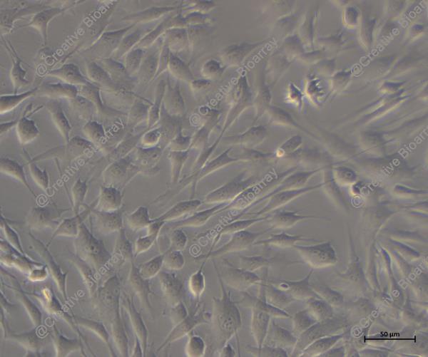
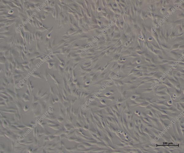
Immortalized Human Colonic Epithelial Cells
Cat.No.: CSC-I1915Z
Species: homo sapiens
Morphology: Polygonal
Culture Properties: Adherent
- Specification
- Background
- Scientific Data
- Q & A
- Customer Review
Note: Never can cells be kept at -20°C.
Researchers derived the Human Colonic Epithelial Cells (HCEC) from healthy adult colonic tissue and achieved cellular immortality by overexpressing CDK4 and hTERT genes through viral vectors. As essential elements of the colonic mucosal barrier colonic epithelial cells protect the intestine from harmful external pathogens. As key elements of the mucosal immune system these cells participate in immune regulation. The HCEC-1CT cell line expresses several stem cell markers such as LGR5, BMI1, CD29, CD44, and Vimentin. The presence of these markers shows that cells can remain undifferentiated yet have the potential to develop into goblet cells, absorptive cells and secretory cells of the colon. Furthermore, these cells possess the ability to express genes related to inflammation such as GBP-1 (Guanylate Binding Protein 1) which become active in inflammatory environments.
HCEC cells serve as a common research model for studying early genetic mutations and signaling pathway abnormalities connected to colorectal cancer because of their benign characteristics. Researchers utilize these cells to study colitis and related intestinal disorders. For example, studies show ANGPTL4 (Angiopoietin-like 4) exerts control over inflammation by regulating chemokines during DSS-induced acute colitis.
Human Colonic Epithelial Cells Exposed to Chronic Inflammation Develop a Resistance Mechanism
Chronic inflammation, as observed in Crohn's disease and ulcerative colitis patients, damages the intestinal mucosa. Collin et al. reasoned that if a subset of intestinal epithelial cells adapt to this inflammatory stress to survive, this adaptation could contribute to their malignant transformation. To explore cellular adaptation mechanisms, they employed human colonic epithelial cells exposed to peroxide or chronic inflammation mimics, combined with bioinformatics analysis of IBD patient datasets and murine carcinogenesis models.
Human primary colonic epithelial cells (HCEC) were immortalized via hTERT transduction (HCEC-hTERT) and cultured with activated macrophage supernatants (AMS, mimicking chronic inflammation) or tert-butyl-hydroperoxide (TBHP, ROS-specific stress) (Fig. 1A). Sublethal doses were applied to induce adaptive responses. After 4-5 days of treatment, cells ceased proliferation (reduced phospho-Serine 10 histone H3, pS10-H3) and entered senescence, marked by acetylated p53 (Ac-p53), p21CIP1 accumulation, and SA-β-galactosidase activity (Fig. 1B). Despite ectopic telomerase regulation, this model recapitulated in vivo observations. Cells maintained senescence for weeks under stress but resumed uniform proliferation after one month, indicating adaptation rather than clonal selection. The surviving populations were termed Esc-Inf (escape from chronic inflammation-induced senescence) and Esc-TBHP (escape from TBHP-induced senescence) (Fig. 1A).
Fig. 1. Human colonic epithelial cells adapt to chronic inflammation-associated oxidative stress in vitro (Collin G, Foy J-P, et al., 2023).
CSE Activity Stimulates TNF-α-Mediated Wound Healing
Cystathionine gamma-lyase (CSE) and TNF-α are now recognized as key regulators of intestinal homeostasis, inflammation, and wound healing. In colonic epithelial cells, both molecules have been shown to influence a variety of biological processes, but the specific interactions between intracellular signaling pathways regulated by CSE and TNF-α are poorly understood.
In the present study, Arroyo Almenas et al. investigated these interactions in normal colonocytes and an organoid model of the healthy human colon using CSE-specific pharmacological inhibitors and siRNA-mediated transient gene silencing in analytical and functional assays in vitro. They demonstrated that CSE and TNF-α mutually regulated each other's functions in colonic epithelial cells. TNF-α treatment stimulated CSE activity within minutes and upregulated CSE expression after 24 h, increasing endogenous CSE-derived H2S production. In turn, CSE activity promoted TNF-α-induced NF-ĸB and ERK1/2 activation but did not affect the p38 MAPK signaling pathway. To explore CSE's role in intestinal repair, they conducted a 2D wound scratch assay with TNF-α (10 ng/mL). TNF-α notably enhanced the wound closure ability of human colonic epithelial cells (HCECs) within 24 hours (Fig. 2A and B). When cells were pretreated with CSE inhibitors PAG or BCA (3 mM), both basal migration and TNF-α stimulation were inhibited (Fig. 2B). In contrast, blocking ERK or p38 MAPK with PD or SB affected only the TNF-α response, as ERK inhibition notably reduced TNF-α-stimulated closure, while p38 did not (Fig. 2B). These findings indicate that TNF-α-induced epithelial healing depends on CSE/H2S activity and ERK1/2 activation.
Fig. 2. CSE activity is indispensable for basal and TNF-α-mediated HCEC wound healing (Arroyo Almenas F; Törő G, et al., 2024).
These are human colonic epithelial cells that have been immortalized to enable continuous proliferation while maintaining essential characteristics of normal colonic epithelial cells. They serve as a robust model for studying various aspects of intestinal biology and disease.
These cells are versatile and can be used in several research areas, including:
Gastrointestinal physiology and pathophysiology.
Colorectal cancer studies.
Drug absorption and metabolism research.
Intestinal barrier function and permeability studies.
Investigations of inflammatory bowel diseases (IBD).
Ask a Question
Write your own review