Rat Periodontal Ligament Fibroblasts
Cat.No.: CSC-C5145S
Species: Rat
Source: Periodontal Ligament; Periodontium
Cell Type: Fibroblast
- Specification
- Background
- Scientific Data
- Q & A
- Customer Review
Rat Periodontal Ligament Fibroblasts from Creative Bioarray are isolated from the rat eye tissue. The method we use to isolate Rat Periodontal Ligament Fibroblasts was developed based on a combination of established and our proprietary methods. The Rat Periodontal Ligament Fibroblasts are characterized by immunofluorescence with antibodies specific to vimentin or fibronectin. Each vial contains 0.5x10^6 cells per ml and is delivered frozen.
Rat periodontal ligament fibroblasts (PLFs) are primary fibroblasts from rat periodontal ligament tissue. The periodontal ligament is a fibrous connective tissue around the root of a tooth. The tooth connects to the alveolar bone through cementum attachment. PLFs are the major cell population in the periodontal ligament. They have a spindle-shaped or stellate flat appearance. PLFs can produce and release some extracellular matrix (ECM) components such as collagen, elastin, and glycoproteins. These cells have been proven to participate in the repair and regeneration of the periodontal ligament. Furthermore, PLFs can phagocytize foreign substances to remove pathogens and metabolic products in the periodontal ligament. PLFs play a crucial role when detecting mechanical forces that act upon the periodontal ligament. Research suggests that PLFs regulate the synthesis and degradation of ECM proteins in response to mechanical stimulation, thus playing a role in the remodeling of the alveolar bone. PLFs can also be induced to differentiate into osteoblasts or odontoblasts, which indicates that PLFs have great value in periodontal tissue regeneration and bone tissue engineering.
Hyaluronidase Inhibits TGF-Β-Mediated Rat Periodontal Ligament Fibroblast Expression of Collagen and Myofibroblast Markers
Orthodontic treatment relies on periodontal tissue remodeling but faces challenges in maintaining retention due to fibrous tissue effects. Collagen fibers and myofibroblasts may stimulate tooth movement recurrence. Hyaluronic acid (HA) is a key component in periodontal tissues, and its degradation by hyaluronidase (HYAL) may influence collagen production and myofibroblast transformation.
This study examines if HYAL can inhibit collagen fiber production and myofibroblast marker expression in rat periodontal ligament cells (rPDLCs) without affecting bone mineralization. Primary rPDLCs, obtained via collagenase digestion and tissue culture, showed triangular and spindle shapes, aligning with pervious study. The culture medium was refreshed every 3 days, and proliferation increased after 7 days, reaching 70–80% confluence by day 10, suitable for passage. At the second generation, cells presented stable morphology and confluence led to radial or vortex arrangements (Fig. 1a). To find the right concentration for experiments, rPDLCs were incubated with varying HYAL concentrations for 1–5 days. HYAL concentrations over 20 U/mL significantly inhibited rPDLC proliferation (20 U/mL: P < 0.05, 100 U/mL: P < 0.01, 500 U/mL: P < 0.001 on day 1; 100 U/mL: P < 0.001, 500 U/mL: P < 0.001 on day 5), while 0.8 and 4 U/mL had no significant effect from days 1 to 5 (Fig. 1b–c). To see if HYAL affects orthodontic recurrence by inhibiting bone mineralization, we analyzed mineralized nodules using Alizarin red staining after treatment with 4 and 20 U/mL HYAL. With 4-MU as the positive control, results indicated that HYAL increased cell gaps and aggregation into clusters. The 20 U/mL group showed more pronounced aggregation than the 4 U/mL group, where mineralized crystals were fully clumped. However, no significant difference in cell mineralization was observed in the 4 U/mL group compared to control (Fig. 1d).
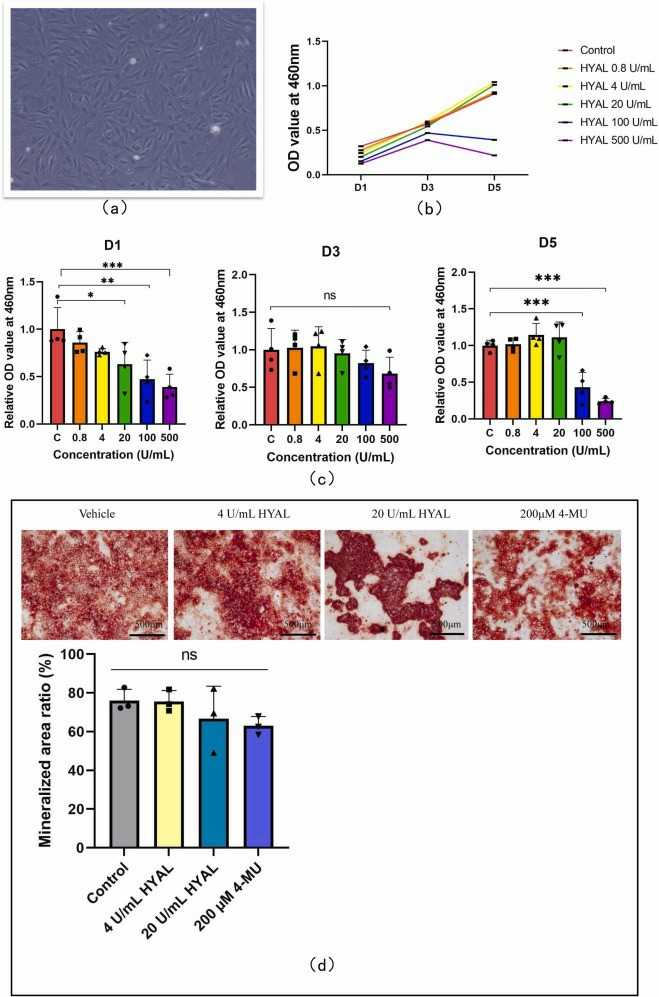 Fig. 1. Analysis of cell proliferation and osteogenic differentiation (Li J, Chen C, et al., 2024).
Caspase 3 Activity in Isolated Fetal Rat Lung Fibroblasts and Rat Periodontal Ligament Fibroblasts: Cigarette Smoke Induced Alterations
Smoking during pregnancy poses risks to the fetus and may cause infant mortality. The oral cavity and its cells are the first to be exposed to cigarette smoke, potentially spreading toxins to other organs. Apoptosis, regulated by caspases, is crucial for embryogenesis and homeostasis. Deregulation of apoptosis is linked to abnormal fetal lung development and adult diseases.
In this study, fetal rat lung fibroblasts and adult rat periodontal ligament fibroblasts were exposed to different concentrations of cigarette smoke extract. The main executioner caspase to be activated in the process of apoptosis is caspase-3. To further analyze Caspase-3 activity in isolated fetal rat lung fibroblasts and PDL fibroblasts exposed to CSE (5%, 10%, and 15%) (v/v) for 60 minutes a fluorometric assay kit was used. Exposure to CSE at concentrations of 10% and15% (v/v) produced significantly elevated activity (p < 0.05) of caspase-3 compared to the non-exposed cells (Fig. 2). No significant differences were observed in the caspase-3 activity in cells exposed to 5% CSE when compared to the non-exposed cells. Interestingly, no significant differences were observed in the caspase-3 activity in PDL fibroblasts (Fig. 3) exposed to 5%, 10% or 15% (v/v) CSE compared to the non-exposed cells.
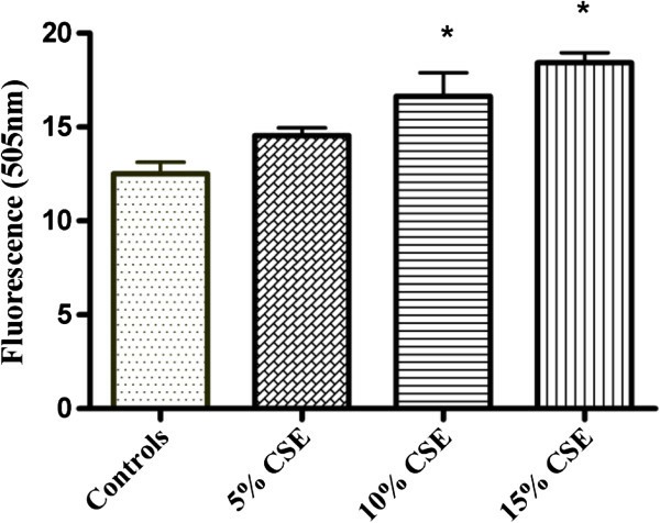 Fig. 2. Effect of CSE on caspase 3 activity in fetal rat lung fibroblasts (Ahmed A, Thliveris J A, et al., 2016).
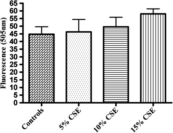 Fig. 3. Effect of CSE on caspase 3 activity in rat periodontal ligament fibroblasts (Ahmed A, Thliveris J A, et al., 2016).
Ask a Question
Write your own review
- You May Also Need

Description: The thoracic aorta is located in the chest cavity and gives off arteries that branch to the esophagus, pericardium, lungs, and trachea. The thoracic aorta can be subdivided into the ascending aorta, ...

Description: Rat Podocytes are isolated from normal rat kidney. The cells are characterized by immunofluorescence with antibodies specific to podocin, Ang1, Nephrin, ACTN4, NPHS2. T25 flasks is required for cell ...

Description: Rat Bronchial Smooth Muscle Cells are isolated from normal rat bronchi tissue. Rat Bronchial Smooth Muscle Cells are characterized by immunofluorescence with antibodies specific to alpha-actin. T25 ...

Description: Guinea Pig Endothelial Cells from Creative Bioarray are isolated from guinea pig tissue. Prior to shipping, cells at passage 2 are detached from flasks and immediately cryopreserved in vials. Each ...

Description: Rat Lung Epithelial Cells are isolated from normal rat lung tissue. The cells are characterized by immunofluorescence with antibodies specific to CK-18, CK-19. T25 flasks is required for cell ...

Description: Rat Vein Endothelial Cells from Creative Bioarray are isolated from inferior vena cava tissue of 6-8 week old laboratory Sprague-Dawley rat. Rat Vein Endothelial Cells are grown in T75 tissue culture ...