
HSC-3
Cat.No.: CSC-C6335J
Species: Homo sapiens (Human)
Source: Lymph Node Metastasis
Morphology: epithelial-like
Culture Properties: Adherent cells
- Specification
- Background
- Scientific Data
- Q & A
- Customer Review
Store in liquid nitrogen.
HSC-3 is an established human oral squamous cell carcinoma (OSCC) cell line derived from a metastatic lymph node of tongue squamous cell carcinoma patient. As mentioned above, because HSC-3 cells were derived from a metastatic tumor site they represent an aggressive OSCC phenotype and have been commonly used in vitro to study mechanisms of invasion, metastasis and tumor progression associated with head and neck cancers.
HSC-3 cells readily propagate as adherent epithelial like monolayers and have polygonal morphology with high proliferation rates. They have increased migratory and invasive capabilities compared to less aggressive OSCC lines in vitro. HSC-3 cells are positive for epithelial markers including cytokeratins. However, they also exhibit changes in cell-cell adhesion and cytoskeletal organization that are associated with malignant transformation.
On a molecular level HSC-3 cells exhibit dysregulation of signaling pathways involved in epithelial-mesenchymal transition (EMT), extracellular matrix degradation, and cell survival (increased expression of matrix metalloproteinases and pro-invasive signaling cascades). Because of these properties HSC-3 cells have been used widely for mechanistic studies examining tumor invasion, metastasis and therapeutic resistance.
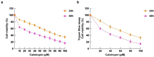
Effect of Calotropin on the Viability of HSC-3 Cells
Calotropin is a cardiac glycoside isolated from Calotropis gigantea has shown promising results in anti-tumorigenesis. Jayaraman et al. aimed to assess the anti-cancer potential of calotropin on HSC-3 oral squamous cancer cells and to understand the mechanism involved. Cytotoxic potential of calotropin against oral cancer HSC-3 cells was analyzed by MTT assay after treating with 10-100 μM for 24 and 48 h. Calotropin significantly caused a reduction in cell viability in dose-dependent manner with higher potency at 48 h (IC50 = 27.53 μM) as compared to 24 h (IC50 = 61.17 μM) treatment (Fig. 1a). Results were further validated by Trypan blue exclusion assay (Fig. 1b), confirming the potent cytotoxic nature of calotropin on HSC-3 cells and its therapeutic effectiveness against oral cancer.
Ask a Question
Write your own review
- You May Also Need
Description: Human cell line derived from oral squamous cell carcinoma occurred in 69-yo, male patient. HLA-A 24/.

Description: Human cell line derived from metastasis of cancer occurred in oral cavity, the same patient as T3M-1 Clone2 and T3M-1 Cl-10.

Description: G-CSF and IL-1 producing oral squamous cell carcinoma, the same patient as T3M-1 Clone2 and CJM.

Description: The CAL-27 cells are established from the poorly differentiated squamous cell carcinoma of the tongue removed from a 56-year-old man before treatment in 1982. CAL 27 cells are epithelial, polygonal ...
Description: Established from the surgically removed fragment of a tongue lesion from a 69-year-old man with moderately differentiated squamous cell carcinoma of the tongue in 1983 (prior to therapy)

- Adipose Tissue-Derived Stem Cells
- Human Neurons
- Mouse Probe
- Whole Chromosome Painting Probes
- Hepatic Cells
- Renal Cells
- In Vitro ADME Kits
- Tissue Microarray
- Tissue Blocks
- Tissue Sections
- FFPE Cell Pellet
- Probe
- Centromere Probes
- Telomere Probes
- Satellite Enumeration Probes
- Subtelomere Specific Probes
- Bacterial Probes
- ISH/FISH Probes
- Exosome Isolation Kit
- Human Adult Stem Cells
- Mouse Stem Cells
- iPSCs
- Mouse Embryonic Stem Cells
- iPSC Differentiation Kits
- Mesenchymal Stem Cells
- Immortalized Human Cells
- Immortalized Murine Cells
- Cell Immortalization Kit
- Adipose Cells
- Cardiac Cells
- Dermal Cells
- Epidermal Cells
- Peripheral Blood Mononuclear Cells
- Umbilical Cord Cells
- Monkey Primary Cells
- Mouse Primary Cells
- Breast Tumor Cells
- Colorectal Tumor Cells
- Esophageal Tumor Cells
- Lung Tumor Cells
- Leukemia/Lymphoma/Myeloma Cells
- Ovarian Tumor Cells
- Pancreatic Tumor Cells
- Mouse Tumor Cells