
Immortalized Porcine Aortic Endothelial Cells (AOC)
Cat.No.: CSC-I9192L
Species: Mus musculus
Source: Aorta
Morphology: Cobblestone-like
Culture Properties: Adherent
- Specification
- Background
- Scientific Data
- Q & A
- Customer Review
Note: Never can cells be kept at -20 °C.
CIK-HT003 HT® Lenti-SV40T Immortalization Kit
2) 51Cr release assay was used to analyse the susceptibility of AOC to xenogeneic cell-mediated cytotoxicity;
3) 1,1’-dioctadecyl-3,3,3’,3’-tetramethylindocarbocyanine (DiI) -labelled Ac-LDL was used to evaluate Ac-LDL update by the AOC.
Immortalized Porcine Aortic Endothelial Cells (AOC) are a spontaneously immortalized cell line obtained from aortic endothelium of a Large White pig. Primary endothelial cells have a limited ability for proliferation, however this cell line was generated via serial passage over a lengthy period until continuous proliferation was achieved, giving it a stable and scalable model for vascular research.
In vitro, the AOC cells maintain the morphological and functional features of artery endothelial cells and create a homogeneous monolayer of polygonal cells. They express major endothelium markers including von Willebrand Factor (vWF), CD31 and VE-Cadherin and retain physiological capabilities including thrombin-induced prostacyclin production. They are cultured in a regular basal medium with added serum under conventional conditions (37°C, 5% CO2).
Because of their porcine origin and human-like cardiovascular physiology, AOC cells are frequently employed in xenotransplantation research, especially in the study of endothelial activation and molecular causes of acute vascular rejection. They also provide a powerful platform to study atherosclerosis, thrombosis, vascular inflammation and shear stress on endothelial biology.
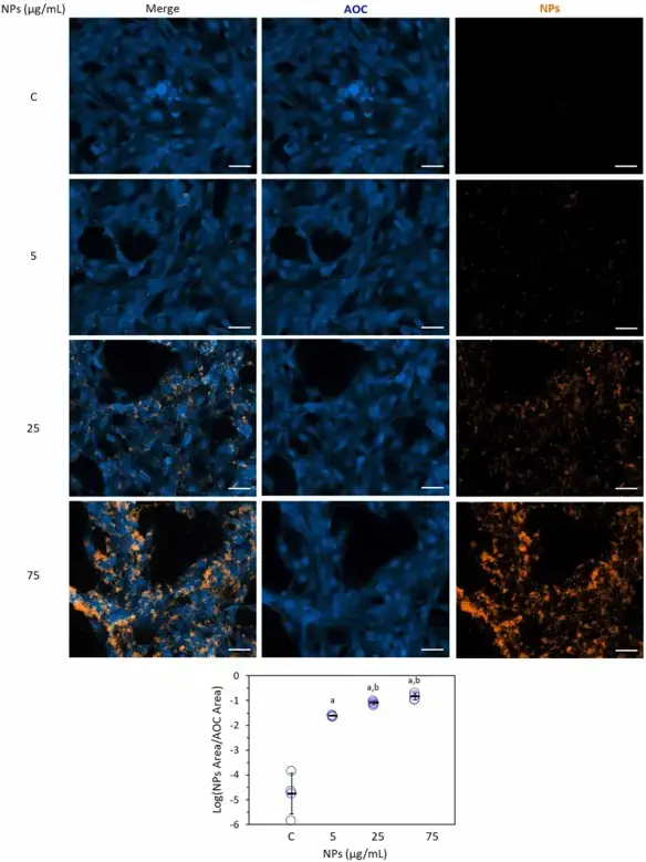
Nanoparticle Interaction with Aortic Endothelial Cells Stimulates VEGF Secretion
Since several research underlined an increased cardiovascular risk due to NPs, present study was undertaken to investigate their effect on aortic endothelial cells (AOC).
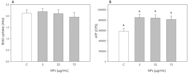
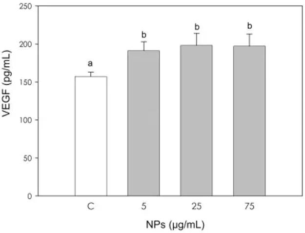
Fluorescence microscopy revealed that NPs specifically colocalized with AOCs at all tested concentrations (5-75 µg/mL), with uptake increasing significantly at higher doses (25 and 75 µg/mL) compared to the lowest concentration (Fig. 1). While NP treatment did not alter cell proliferation (Fig. 2A), it significantly stimulated metabolic activity, as measured by ATP production (Fig. 2B). Similarly, VEGF secretion was significantly increased by all NP concentrations (Fig. 3); however, this effect occurred independently of changes in VEGFgene expression (Fig. 4). These findings suggest that NPs can directly interact with endothelial cells and modulate their secretory function.

Ask a Question
Write your own review

Description: Immortalized Cynomolgus Monkey Pulmonary Artery Smooth Muscle Cells-GFP provided by Creative Bioarray have been developed by immortalizing cynomolgus monkey pulmonary artery smooth muscle cells with ...
Description: Immortalized Monkey Spleen Fibroblasts-SV40T were developed from monkey tissues transduced with a lentiviral expression vector containing the SV40T gene. The cell line was continuously cultured for ...
Description: Immortalized Monkey Bronchial Fibroblasts-GFP provided by Creative Bioarray have been developed by immortalizing monkey bronchial fibroblasts with SV40 Large T antigen and transfecting with tGFP. The ...

Description: Immortalized Canine Kidney Fibroblasts-SV40 have been obtained immortalizing Canine Kidney Fibroblasts with Lenti-SV40 Lentivirus. Immortalized cells were controlled passaging side by side with the ...
Description: Immortalized Monkey Primary Aortic Endothelial Cells-GFP provided by Creative Bioarray have been developed by immortalizing primary monkey aortic endothelial cells with SV40 Large T antigen and ...
Description: Immortalized Monkey Pancreatic Epithelial Cells-SV40T were developed from monkey tissues transduced with a lentiviral expression vector containing the SV40T gene. The cell line was continuously ...
- Adipose Tissue-Derived Stem Cells
- Human Neurons
- Mouse Probe
- Whole Chromosome Painting Probes
- Hepatic Cells
- Renal Cells
- In Vitro ADME Kits
- Tissue Microarray
- Tissue Blocks
- Tissue Sections
- FFPE Cell Pellet
- Probe
- Centromere Probes
- Telomere Probes
- Satellite Enumeration Probes
- Subtelomere Specific Probes
- Bacterial Probes
- ISH/FISH Probes
- Exosome Isolation Kit
- Human Adult Stem Cells
- Mouse Stem Cells
- iPSCs
- Mouse Embryonic Stem Cells
- iPSC Differentiation Kits
- Mesenchymal Stem Cells
- Immortalized Human Cells
- Immortalized Murine Cells
- Cell Immortalization Kit
- Adipose Cells
- Cardiac Cells
- Dermal Cells
- Epidermal Cells
- Peripheral Blood Mononuclear Cells
- Umbilical Cord Cells
- Monkey Primary Cells
- Mouse Primary Cells
- Breast Tumor Cells
- Colorectal Tumor Cells
- Esophageal Tumor Cells
- Lung Tumor Cells
- Leukemia/Lymphoma/Myeloma Cells
- Ovarian Tumor Cells
- Pancreatic Tumor Cells
- Mouse Tumor Cells