QualiCell® Human Amniotic Fluid Stem Cells
- Specification
- Background
- Scientific Data
- Q & A
- Customer Review
Human Amniotic Fluid Stem Cells (hAFSCs) are fetal-derived stem-like cells derived from human amniotic fluid which is most commonly collected from the mother's womb during amniocentesis as part of routine prenatal screening. The cells within the amniotic fluid consist of a variety of different cell types present within the fetus and only represent a small subset of cells within the fluid demonstrate stem cell characteristics including surface expression of the cell surface marker CD117 (c-kit).
hAFSCs represent a cell type that is intermediate between embryonic stem cells and adult stem cells, specifically adult mesenchymal stem cells. They have an extensive proliferative capacity and can be expanded in vitro for several passages while maintaining genetic stability. They can differentiate into osteogenic, adipogenic, chondrogenic, neural, and hepatic lineages when given appropriate conditions. Along with low immunogenicity and low ethical controversy they can be translated easily into various fields of study.
In terms of applications, hAFSCs have shown significant promise in regenerative medicine, where they are used for tissue repair in models of cardiac injury, kidney disease, and neurodegeneration. In tissue engineering, they serve as seed cells for constructing bone, cartilage, and skin substitutes. They are also valuable in prenatal and perinatal therapy, offering potential for early intervention in congenital disorders. Furthermore, hAFSCs are increasingly utilized in disease modeling and drug screening, particularly for studying genetic diseases and developmental biology, due to their fetal origin and stable expansion capacity.
In vitro Evaluation of a Decellularized Human Fetal Skin-Derived Scaffold Repopulated with Human Amniotic Fluid Stem Cells for Potential Application in Myelomeningocele Repair
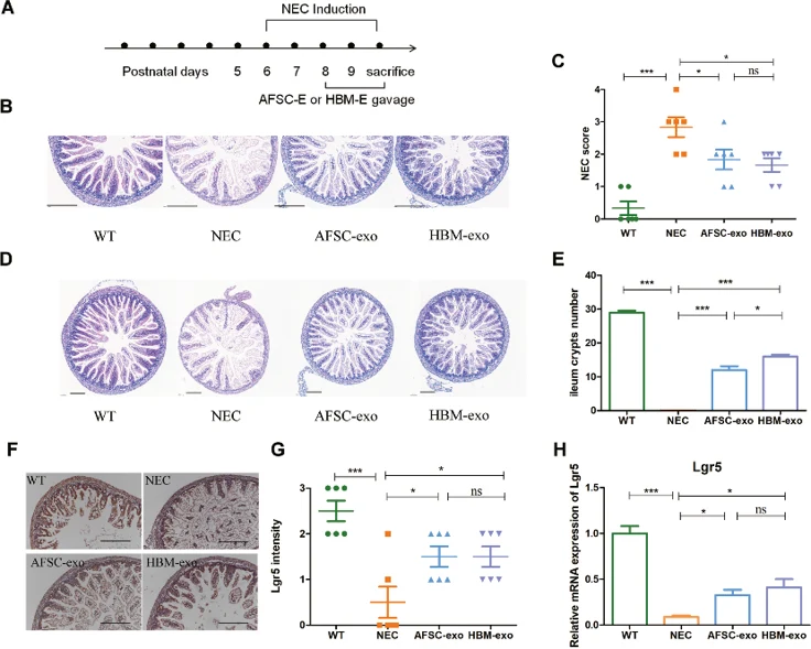
Myelomeningocele (MMC) causes severe lifelong disability through prenatal spinal cord injury. Cavalheiro et al. aimed to develop a decellularized fetal skin scaffold recellularized with human amniotic fluid stem cells (hAFSCs) as a potential regenerative therapy for MMC repair.
Primary cultures were established from all hAF samples (n = 5). After one week with medium changes every other day, hAFSC cultures initially showed heterogeneous populations: epithelioid cells (ECs) (Fig. 1A), fibroblastic cells (FCs) (Fig. 1B), and amniotic fluid-specific cells (AFCs) (Fig. 1C). AFCs and FCs appeared initially, while ECs emerged later and were not present in all samples. After three passages, cultures became homogeneous with fibroblast-like morphology (Fig. 1C), confirmed by scanning electron microscopy (Fig. 1D).
Ultrastructural analysis revealed spindle-shaped cells with cytoplasmic extensions and oval nuclei (Fig. 1E). Dispersed chromatin and visible nucleoli indicated active protein synthesis. The cytoplasm contained well-developed rough endoplasmic reticulum, Golgi complex, and elongated or round mitochondria with defined ridges and vesicles (Fig. 1E, F).
Ask a Question
Write your own review
- You May Also Need
- Adipose Tissue-Derived Stem Cells
- Human Neurons
- Mouse Probe
- Whole Chromosome Painting Probes
- Hepatic Cells
- Renal Cells
- In Vitro ADME Kits
- Tissue Microarray
- Tissue Blocks
- Tissue Sections
- FFPE Cell Pellet
- Probe
- Centromere Probes
- Telomere Probes
- Satellite Enumeration Probes
- Subtelomere Specific Probes
- Bacterial Probes
- ISH/FISH Probes
- Exosome Isolation Kit
- Human Adult Stem Cells
- Mouse Stem Cells
- iPSCs
- Mouse Embryonic Stem Cells
- iPSC Differentiation Kits
- Mesenchymal Stem Cells
- Immortalized Human Cells
- Immortalized Murine Cells
- Cell Immortalization Kit
- Adipose Cells
- Cardiac Cells
- Dermal Cells
- Epidermal Cells
- Peripheral Blood Mononuclear Cells
- Umbilical Cord Cells
- Monkey Primary Cells
- Mouse Primary Cells
- Breast Tumor Cells
- Colorectal Tumor Cells
- Esophageal Tumor Cells
- Lung Tumor Cells
- Leukemia/Lymphoma/Myeloma Cells
- Ovarian Tumor Cells
- Pancreatic Tumor Cells
- Mouse Tumor Cells