Dual-Modality Imaging of IF and IMC
BioRxiv [Preprint]. 2023 Feb 23; 2023.02.23: 529718.
Authors: Kim EN, Chen PZ, Bressan D, Tripathi M, Miremadi A, di Pietro M, Coussens LM, Hannon GJ, Fitzgerald RC, Zhuang L, Chang YH.
INTRODUCTION
Imaging mass cytometry (IMC) is a powerful multiplexed tissue imaging technology that allows simultaneous detection of more than 30 makers on a single slide. It has been increasingly used for single-cell-based spatial phenotyping in a wide range of samples. However, it only acquires a small, rectangle field of view (FOV) with a low image resolution that hinders downstream analysis.
METHODS
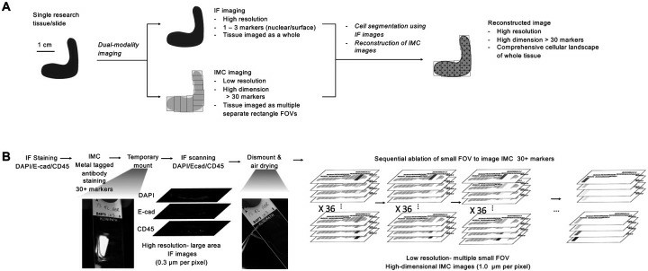
Fig. 1 Experimental benchwork workflow for integrating dual-modality imaging.
- Staining and dual-modality imaging of IF and IMC. Briefly, we stained the slide for two rounds: first with the primary IF antibodies (E-Cadherin, CD45), and second with the secondary IF antibodies and a cocktail of metal isotope-conjugated IMC antibodies (36 markers). The slides were stained with DAPI before temporarily being mounted for whole slide fluorescent scanning for IF markers, followed by dismount and air drying, and acquisition of multiple small rectangle FOVs of IMC.
- We developed a computational approach to map mm2-sized smaller FOV IMC images onto cm2-sized larger WSI IF images, and then register and stitch them together. We successfully registered high-dimensional IMC images with 36 markers onto WSI IF with a high subcellular resolution.
- Once the whole tissue was reconstructed for both IF and IMC, we further registered the image with an adjacent slide stained for hematoxylin and eosin (H&E). We then generated cell masks using MESMER, a deep learning-based algorithm for nuclear and whole-cell segmentation, from the registered high-resolution IF using IF DAPI for the nuclear marker and IF E-Cadherin for the epithelial cell membrane marker.
- Browse our recommendations
Creative Bioarray provides professional products and services, including but not limited to the following.
| Service Types | Description |
| Immunohistochemistry (IHC), Immunofluorescence (IF) Service | Creative Bioarray will give you the best and most comprehensive service in regular and customized immunohistochemistry and immunofluorescence services. |
| Whole Slide Imaging | The whole slide scanning system used by Creative Bioarray provides digital images of the highest available sensitivity and definition and can be used for any tissue and any stain, including H&E, IHC, and ISH. |
| Histological Stains & Dyes | Creative Bioarray provides histological staining methods and related products to help our clients achieve the best dyeing purposes. |
RESULTS
- To demonstrate its applicability in different tissue types, we stained large endoscopic mucosal resection (EMR) tissues of early esophageal adenocarcinoma with adjacent precancerous Barrett’s esophagus, which represents various histologic features, including squamous and glandular epithelia and stromal tissue, and pathological progress from normal tissues to metaplastic precancerous lesion, dysplasia, and cancer in a large area measuring approximately 0.5 cm×2 cm.
- Histomorphologically, the inverted N/C ratio decreases along with disease progression from intestinal metaplasia to dysplasia and adenocarcinoma, which was faithfully recapitulated by the IF-based cell segmentation but not IMC-based, indicating that IF-based segmentation is sufficiently sensitive for subcellular phenotyping, especially for clinical samples.
- Both IF-based and IMC-based cell segmentation showed very low double positivity on two mutually exclusive marker pairs, PanCK/αSMA, and PanCK/CD4. Finally, we evaluated the batch effect in IF and IMC by assessing the signals of the same markers that were acquired on different experiment dates. We found the batch effect was inevitable for IF markers that varied intensities were observed in different batches.
SUMMARY
Here, we reported a highly practical dual-modality imaging method that combines high-resolution IF and high-dimensional IMC on the same tissue slide. Our computational pipeline uses the WSI of IF as a spatial reference and integrates small FOVs IMC into a WSI of IMC. The high-resolution IF images enable accurate single-cell segmentation to extract robust high-dimensional IMC features for downstream analysis.
RELATED PRODUCTS & SERVICES
Reference
- Kim EN, et al. (2023). "Dual-modality imaging of immunofluorescence and imaging mass cytometry for whole slide imaging with accurate single-cell segmentation." bioRxiv. 2023.02.23, 529718.