A Novel Rodent Model of Sporadic Alzheimer's Disease
International Journal of Molecular Science. 2023 Apr 9; 24 (8): 6953.
Authors: Chadwick W, Maudsley S, Hull W, Havolli E, Boshoff E, Hill MDW, Goetghebeur PJD, Harrison DC, Nizami S, Bedford DC, Coope G, Real K, Thiemermann C, Maycox P, Carlton M, Cole SL.
INTRODUCTION
- Sporadic Alzheimer's disease (sAD) is characterized by a progressive loss of cognitive function precipitating from multiple pathologies and is fast becoming an economic burden worldwide. The etiology of sAD is the result of multifaceted interactions among numerous genetic, epigenetic, proteostasis, and environmental factors.
- Almost 95% of current AD patients are associated with sAD as opposed to patients presenting with well-characterized genetic mutations that lead to AD predisposition, i.e., familial AD (fAD). As significant differences in etiology exist between sAD and fAD, it is perhaps more appropriate to develop novel, more sAD-reminiscent experimental models that would expedite the discovery of effective therapies for the majority of AD patients.
METHODS
- Animals. Male C57Bl6/j mice were used throughout the study. Animals were group-housed on a 12 h: 12 h light-dark cycle and food and water were available ad libitum.
- D-Galactose (oDGal) Treatment. D-galactose (2 g/kg or 4 g/kg) and 0.1% sodium benzoate were supplemented into the oDGal group's drinking water; vehicle (Veh) groups received 0.1% sodium benzoate in their drinking water.
- Synaptosome preparation. Hippocampal and cortical synaptosomes were isolated from mice pretreated with vehicle or 4 g/kg oDGal for 8 weeks. The required aliquots of synaptosomal proteins were finally centrifuged at 3020×g/10 min/°C. Supernatants were removed and pellets were stored at 4°C until use in the experiment.
- Mitochondrial functional assay. Mitochondrial function was measured using the Seahorse extracellular flux (XF) 96 analyzer. All experiments were performed at 37°C.
- ATP assay, protein carbonyl, and AGE quantification. These assays were performed using the appropriate kits.
- Locomotor activity assay (LMA), rotarod analyses, novel object recognition (NOR), novel object location (NOL), spatial recognition/topographical test.
- Browse our recommendations
With extensive experience and state-of-the-art technologies, Creative Bioarray is offering Alzheimer's disease modeling and assays to help our customers open the door for generating new insight into disease pathophysiology and improving the process of drug development.
| Service Types | Description |
| Alzheimer's Disease Modeling and Assays | Getting treatment or prevention of AD has attracted extensive attention worldwide. Our services can help our customers open the door to generating new insights into disease pathophysiology. |
| In Vitro Model Service | We offer the development of custom-designed in vitro models by using primary neuronal cultures, cell lines, and iPS cells with genetic modifications. |
| In Vivo Model Service | Animal models of Alzheimer's disease are provided, including but not limited to aging models, transgenic animal models, induced damage models, drosophila models, Caenorhabditis elegans models, and Danio Rerio models. |
| Alzheimer's Disease Study Tools | Our services include but are not limited to behavior tests, neurological imaging, electrophysiology assay, and microdialysis. |
| Alzheimer's disease-related tests | Aβ induced neurotoxicity assay, Amyloid β (Aβ) peptide aggregation inhibition assay, Aβ peptide formation or screening for secretase inhibitors, Tau hyperphosphorylation assay, Tau aggregation assay, Neurite outgrowth assay |
RESULTS
- oDGal significantly impairs basal mitochondrial function in both the hippocampal and the cortical synaptosomes. ATP production is significantly impaired in the hippocampal but not the cortical synaptosomes. Both oDGal groups exhibited a dose-dependent increase in soluble AGE in the cortex and frontal cortex. Elevations in insoluble AGE were also observed in the cortex.
- oDGal treatment significantly increases both insoluble Aβ40 and Aβ42 levels in the cortex, frontal cortex, hippocampus, and cerebellum. Soluble p-tau antibody exposure revealed a broad immunoreactive band extending from 55 kDa to 150 kDa for both oDGal groups in all brain regions except the cerebellum. A considerable inflammatory response in the oDGal treatment groups—the frontal cortex and cortex—displayed the majority of the inflammatory marker response.
- oDGal treatment promotes multi-domain cognitive deficits without affecting motor coordination or activity. In addition to insoluble AGE, PC, and soluble Aβ42, both insoluble Aβ40 and Aβ42 also may harm spatial memory.
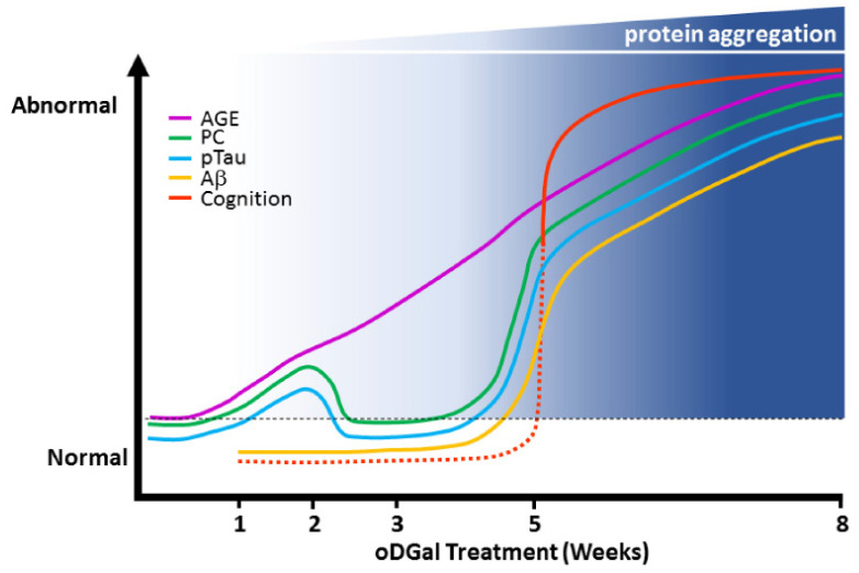 Fig. 1 Diagram illustrating sequential onset of pathologies in the hippocampus following oDGal dosing.
SUMMARY
The study presents the oDGal mouse model, a novel model of sAD that displays a range of AD-like pathologies as well as multiple cognitive deficits reminiscent of AD symptomology.
RELATED PRODUCTS & SERVICES


Reference
- Yau E, et al. (2023). "Investigation of simplified physiologically-based pharmacokinetic models in rat and human." CPT Pharmacometrics Syst Pharmacol. 12 (3), 333-345.