Rat Aortic Ring Assay
Angiogenesis, the sprouting of blood vessels from preexisting vasculature is associated with both natural and pathological processes. Various angiogenesis assays involve studies of individual endothelial cells under culture conditions, such as the Boyden Chamber assay, wound healing assay, and tube formation assay. These assays mainly use monolayers of endothelial cells which are modified by repeated passages and are fully proliferative, a situation far away from physiology. In addition, not only endothelial cells are involved in this process but surrounding cells (such as pericytes, smooth muscle cells, and fibroblasts) and the supporting matrix are also major players.
The three-dimensional in vitro aortic ring model recapitulates the complexities of angiogenesis combining the advantages of in vitro and in vivo models. The aortic rings are cultivated in a chemically defined culture environment. Microvessels grown in this system are lumenized vessels with surrounding supporting cells, which are almost indistinguishable from the microvessels formed during angiogenesis in vivo.
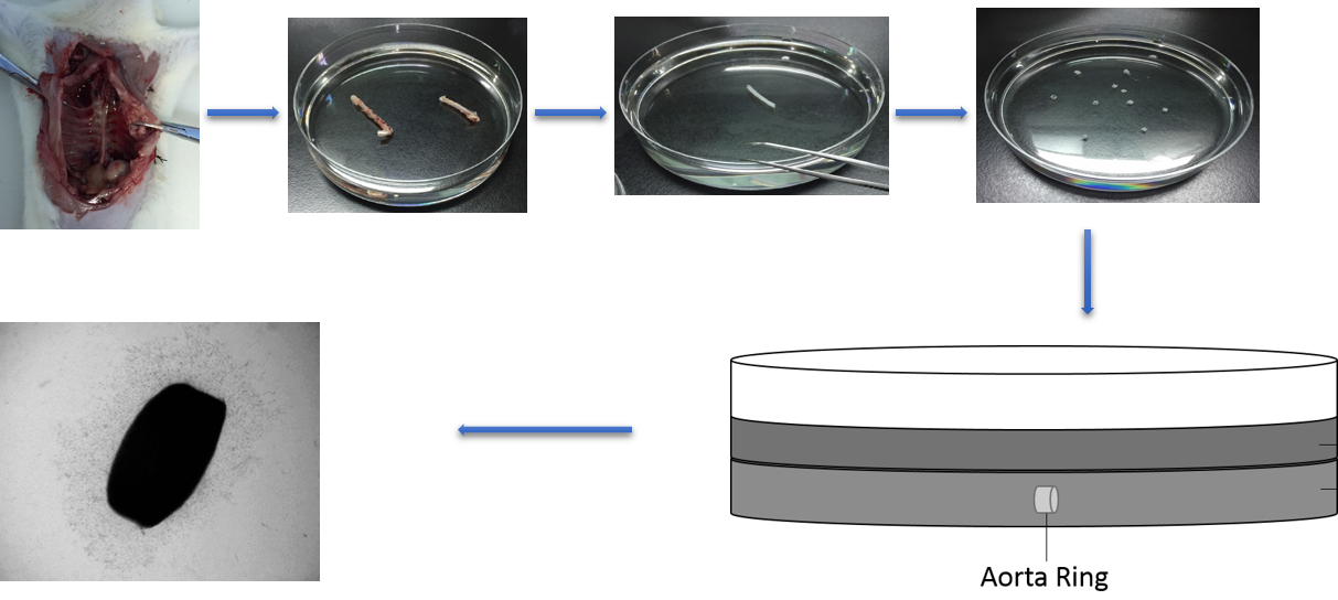 Figure 1. The workflow of rat aortic ring assay.
Materials and Equipment
Growth factor-reduced matrigel
24-well plates
Pipette tips
SD Rat (6-8 weeks)
Chloral hydrate (4%)
Ethanol
Surgical tweezers, scissors and scalpel
Phosphate buffer saline (PBS)
Dissecting microscope
Incubator
DMEM medium
Fetal bovine serum (FBS)
Inverted light microscope
Assay Procedure
All of the following steps will be performed under sterile conditions.
Rat aortic ring preparation
- Allow the growth factor-reduced matrigel melt overnight from -20°C to 4°C in a refrigerator.
- Put 24-well plates and pipette tips in -20°C chilled overnight.
- Inject 4% chloral hydrate to rat’s abdomen and then sacrifice the animal through cervical dislocation.
- Place the rat on a dissecting board and disinfect skin with 80% ethanol.
- Use scissors to open the chest and remove other organs to expose the thoracic aorta.
- Use tweezers and scissors to separate the spine and aorta.
- Cut the aorta from diaphragm to the end of the heart, then put it in a 10 cm dish and wash the aorta with sterile PBS at room temperature.
- Carefully clean the surrounding tissue around the aorta under a dissecting microscope.
- When the aorta is clean, transfer it to a clean dish and keep it on ice.
- Cross-section the thoracic aorta into rings that are 1 to 2 mm in length (20 to 25 rings/aorta) using a scalpel.
Aortic rings embedding
- Homogenously add 200 µL of matrigel into the bottom of each well in a pre-chilled 24-well plate using pre-chilled tips.
Note: 1) The matrigel should be added carefully and quickly because it is very easy to solidify at room temperature. 2) After adding the matrigel, gently shake the plate to ensure that the whole surface is covered by the matrigel and there are no bubbles in the well. - Put the plate into a CO2 incubator (37°C) for 30 min to allow polymerization.
- Place the rings in the middle of each well.
- Add another 200 µL matrigel into the well to cover the rings.
- Place the plate back in the incubator and incubate for 30 min.
- After 30 min, add 500 µL of DMEM medium supplemented with 10% FBS per well, finally put the 24-well plate in the incubator.
Quantification
- Refresh the medium every 2 d.
- After 7 d, the branch of vascular is observed under microscope.
- Use inverted light microscope to detect the sprouting length and range.
References
- Baker M, et al.; Use of the mouse aortic ring assay to study angiogenesis. Nature Protocols, 2012, 7: 89-104.
- Bellacen K, et al.; Aortic ring assay. Journal of Visualized Experiments, 2009, 33: 1564.
Cell Services:
Cell Line Testing and Assays