Establishment Protocol for Nude Mouse Models of Pancreatic Cancer
GUIDELINE
Pancreatic cancer is a tumor with high malignancy, rapid development, and extremely poor prognosis. Early symptoms are atypical, and by the time obvious symptoms appear, it has already entered the advanced stage and lost the chance of radical treatment. It is not sensitive to traditional radiotherapy and chemotherapy, and even for patients who have successfully undergone surgery, the 5-year survival rate is only close to 20% due to easy metastasis after surgery.
To study the biological characteristics of pancreatic cancer and explore the effective measures for its diagnosis and treatment, it is necessary to establish animal models. In this experiment, subcutaneous implantation was used, the biological characteristics of tumor cells remained intact, the light and electron microscopic observation of tumor tissues conformed to the original structural characteristics of cell line 8988, and there was no obvious difference in the morphology, and the morphology of tumor tissues in cell culture conformed to that of cell line 8988, and the differences between animals were small, and the serum test did not find any tumor-associated antigens. infiltration, no remote metastasis caused.
METHODS
Preparation of experimental materials
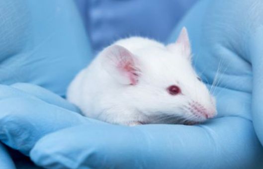
- Nude mice BALB/ c nu- nu, 4-6 weeks old, weighing 16-20 g, both male and female, were kept in an SPF grade environment with a constant temperature of 25-27°C, a constant humidity of 45%-50%, and drinking water and food were sterilized.
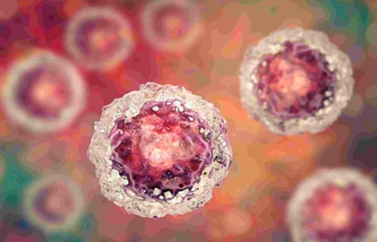
Human pancreatic cancer cell line 8988 in vitro subculture
- Put the human pancreatic cancer cell line into RPMI-1640 complete medium containing 10% of RPMI-1640, put it in 37°C, 5% CO2 incubator, change the liquid every 2 d, 0.25% trypsin digestion, a passaging 3 passaging culture.
- When the cells filled the bottom of the bottle, a single cell suspension was collected, 5 × 10⁶ tumor cells per cell, 0.2 ml, injected into the axilla of nude mice subcutaneously to establish a tumor model.
- Tumor size was measured weekly with vernier calipers and calculated according to the formula V = π / 6 × length × width × height, and the growth curve was plotted.
Serology
- At the 6th week, mice were executed by eye-pulling method, blood was collected in test tubes, serum was extracted from the test tubes, and tumor-associated antigen and amylase were detected.
Giant test
- After the nude mice were executed, the tumor was peeled off subcutaneously, and the texture, infiltration, and blood supply were recorded, and the presence or absence of abdominal adhesions and the metastatic status of each organ were observed.
- Tumor tissue, lung, liver, spleen, mesentery, duodenum, stomach, brain, and other specimens were taken and placed in 10% formalin fixative for use.
Pathologic examination
- The above specimens were routinely embedded in paraffin, stained with HE staining, and observed by light microscope.
Ultrastructure observation
- Tumor tissues were double-fixed with 2.5% glutaraldehyde and 1% osmium acid, embedded, ultrathin sectioned, double-stained with uranium and lead, and observed by transmission electron microscope.
Microscopic examination
- Tumor tissue cell culture and live cell observation. Take fresh subcutaneous tumor tissue from nude mice, rinse it with PBS 3 times, cut it into 1 m3 size, digest it with 0.25% trypsin, remove the coarse tissue, and then make a single-cell suspension, place it in 37°C, 5% CO2 incubator for culture, observe its morphology and growth under an inverted microscope, and compare it with cell line 8988.
- HE staining and morphological observation of cells. Tumor cells and cell lines were cultured in the same amount in six-well plates containing coverslips. After growth to secondary fusion, the medium was discarded, washed three times with PBS, fixed with 2.5% glutaraldehyde, washed three times with PBS, and then stained with HE, and observed and compared with those of cell line 8988 under the light microscope.
Creative Bioarray Relevant Recommendations
- Creative Bioarray is a renowned biological company with extensive experience in providing high-quality pancreatic tumor cells for research purposes.
| Cat. No. | Product Name |
| CSC-C0307 | PA-TU-8988T |
| CSC-C0312 | PA-TU-8902 |
| CSC-C0326 | PA-TU-8988S |
| CSC-C0333 | HUP-T4 |
| CSC-C0359 | HUP-T3 |
| CSC-C0430 | YAPC |
View the details of our pancreatic tumor cells and find what you need!
- We provide a series of histological staining methods and related products to help our clients achieve the best dyeing purposes. In addition, we perform full comprehensive transmission electron microscopy (TEM) service for the biological sciences and clinical research including plant samples, animal samples, bacteria, and pathology specimens.
NOTES
- Different experimental groups and control groups should be designed, and the number of animals in each group is usually 5-10.
- Generally speaking, after 6-8 weeks of inoculation, when the tumors of mice can grow to 15-20 mm in diameter (i.e. the mice will be on the verge of death), blood can be taken from the eyeballs, serum can be separated and preserved, and then the mice can be executed, and the tumor tissues can be photographed, and the tumor tissues can be taken as frozen tissue sections (or preserved at -80°C) for corresponding immunohistochemistry staining, and some of the tumor tissues can be fixed with 3% neutral formaldehyde, embedded in paraffin for routine HE staining, and then embedded in paraffin for routine HE staining. Some tumor tissues were fixed with 3% neutral formaldehyde, embedded in paraffin, and stained with conventional HE staining.