Preparation Protocol for FISH Probes
GUIDELINE
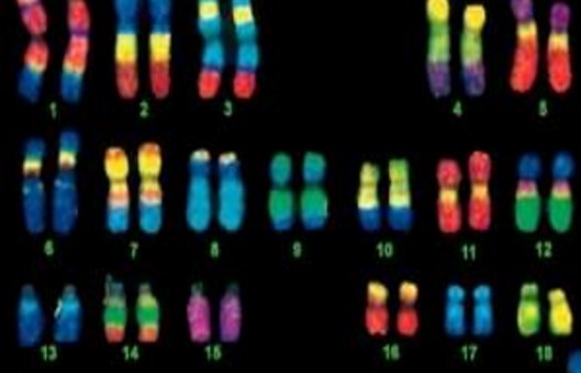
- Fluorescence in situ hybridization (FISH) began with the combination of traditional cytogenetic and DNA techniques.
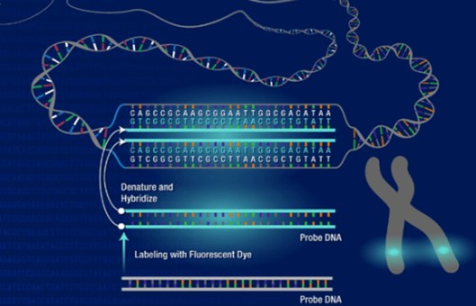
- It is based on the Southern blot principle, where a known nucleic acid molecule indirectly labeled with a semi-antigen such as biotin, digoxin or directly labeled with fluorescein is used as a probe. The probe and target sequence double-stranded DNA is denatured and then hybridized. The complementary heterologous single-stranded DNA molecules are annealed at a suitable temperature and ionic strength to form stable heterologous double-stranded DNA, the semi-antigens are displayed by fluorescently labeled affinity or anti-digoxin antibodies, and the hybridization signal is observed by fluorescence microscopy.
METHODS
Notch translation marker probes
- The notch panning system for 1μg DNA is 0.1 mmol/L dTTP 6.5 μl, 0.1 mmol/L dNTP 10 μl, 10×Nick Translation buffer 5 μl, 1 mmol/L DIG-11-dUTP /Biotin-16-dUTP 0.5 μl, Nick Translation Enzyme 10 μl, DNA (1μg) X μl, and H2O 18-X μl. For plasmids ≤10kb, react at 15°C for 3.5 hours; for BAC/PAC, react at 15°C for 9 hours.
- Place the EP tube on ice, take 5 μl of it, and heat it at 70°C for 5 minutes, then perform 2% agarose gel electrophoresis to observe the size of the labeled probe, the suitable size of the single sequence probe is 200-600 bp; if the fragment size is large, extend the reaction at 15°C for 10-30 minutes, then repeat the gel electrophoresis until the suitable size of the probe is obtained.
Precipitation and denaturation of probes
- Composition of the probe mixture. For BAC/PAC probes, DNA probe 5 μl, Human Cot-1 DNA 3 μl, Salmon Sperm DNA 0.5 μl, H2O 1.5 μl. For mitotic probes, DNA probe 5 μl, Salmon Sperm DNA 0.5 μl, H2O 4.5 μl.
- Precipitation of DNA. The probe mixture was mixed with 1 μl (V/10) 3M sodium acetate (pH 5.2) and 27.5 μl (2.5V) anhydrous ethanol (lyophilized at -20°C), precipitated at -80°C for 30 min (or -20°C overnight), and centrifuged at 14,000 g at 4°C for 30 min to precipitate DNA.
- Wash the precipitate. Carefully discard the supernatant, wash once with 70% ethanol, and centrifuge again at 14,000 g for 15 min at 4°C; carefully discard the supernatant and air-dry the DNA precipitate in a medium temperature water bath at 45-50°C for 10-15 min.
- Dissolve the probe. Add 5 μl of pre-warmed deionized formamide (pH 7.0) at 37°C, centrifuge for a short time and shake at 37°C for 30 min to fully dissolve the DNA; then add 5 μl of pre-warmed Master Mix at 37°C, centrifuge for a short time and shake at 37°C for 15-30 min.
- Denaturation and pre-hybridization of probes. Short centrifugation followed by denaturing probes in an 80°C water bath for 10 min and an ice bath for 5 min; short centrifugation and pre-hybridization in a 37°C water bath for 30-60 min; unique sequence probes (e.g. cDNA) and repeat sequence probes (e.g. α-satellite DNA) probes, no pre-hybridization is required.
- Hybridize at 37°C overnight in a humidity chamber.
Creative Bioarray Relevant Recommendations
- Creative Bioarray provides the most comprehensive list of FISH probes for the rapid identification of a wide range of chromosomal aberrations across the genome. We continue to expand the scope of the FISH probes to meet the customer's research needs- including the introduction of Customized FISH Probes.
NOTES
- When most of the ethanol evaporates, the white DNA precipitate becomes translucent; be sure to remove the ethanol thoroughly, otherwise, tiny precipitates will be generated after the addition of Master Mix, resulting in high background.
- The DNA precipitate can also be dissolved in TE buffer 1.1 μl and diluted by adding Vysis probe buffer 4.4 μl, mixed and centrifuged instantaneously, and shaken for 15-30 min at 37°C.
RELATED PRODUCTS & SERVICES
For research use only. Not for any other purpose.
Resources
- FAQ
- Posters & Downloads
- Protocol
- Cell Culture Guide
- Technical Bulletins
-
Explore & Learn
-
Cell Biology
- Monocytes vs. Macrophages
- How to Detect and Remove Endotoxins in Biologics?
- Comparison of Different Methods to Measure Cell Viability
- What Are Myeloid Cell Markers?
- How to Start Your Culture: Thawing Frozen Cells
- Biomarkers and Signaling Pathways in Tumor Stem Cells
- Techniques for Cell Separation
- Circulating Tumor Cells as Cancer Biomarkers in the Clinic
- CFU Assay for Hematopoietic Cell
- Comparison of the MSCs from Different Sources
- T Cell Activation and Expansion
- How to Isolate and Analyze Tumor-Infiltrating Leukocytes?
- Contamination of Cell Cultures & Treatment
- Generation and Applications of Neural Stem Cells
- Stem Cell Markers
- Cell Cryopreservation Techniques and Practices
- Guidelines for Cell Banking to Ensure the Safety of Biologics
- Critical Quality Attributes and Assays for Induced Pluripotent Stem Cells
- What Is Cell Proliferation and How to Analyze It?
- Direct vs. Indirect Cell-Based ELISA
- Comparison of Several Techniques for the Detection of Apoptotic Cells
- STR Profiling—The ID Card of Cell Line
- How to Assess the Migratory and Invasive Capacity of Cells?
- Cryopreservation of Cells Step by Step
- What are PBMCs?
- Quantification of Cytokines
- What Cell Lines Are Commonly Used in Biopharmaceutical Production?
- Neural Differentiation from Induced Pluripotent Stem Cells
- Isolation, Expansion, and Analysis of Natural Killer Cells
- Tumor Stem Cells: Identification, Isolation and Therapeutic Interventions
- Cell Culture Medium
- IL-12 Family Cytokines and Their Immune Functions
- Multi-Differentiation of Peripheral Blood Mononuclear Cells
- How to Scale Up Single-Cell Clones?
- What are Mesothelial Cells?
- T Cell, NK Cell Differentiation from Induced Pluripotent Stem Cells
- Major Problems Caused by the Use of Uncharacterized Cell Lines
- What are the Differences Between M1 and M2 Macrophages?
- Mesenchymal Stem Cells: A Comprehensive Exploration
- Human Primary Cells: Definition, Assay, Applications
- Enrichment, Isolation and Characterization of Circulating Tumor Cells (CTCs)
- Organoid Differentiation from Induced Pluripotent Stem Cells
- Tips For Cell Cryopreservation
- How to Decide Between 2D and 3D Cell Cultures?
- CHO Cell Line Development
- How to Eliminate Mycoplasma From Cell Cultures?
- Troubleshooting Cell Culture Contamination: A Comprehensive Guide
- Unveiling the Molecular Secrets of Adipogenesis in MSCs
- How to Isolate PBMCs from Whole Blood?
- How to Handle Mycoplasma in Cell Culture?
- Strategies for Enrichment of Circulating Tumor Cells (CTCs)
- ddPCR vs qPCR vs NGS: Which Platform Fits Your Research?
- Spheroid vs. Organoid: Choosing the Right 3D Model for Your Research
- From Collection to Cure: How ACT Works in Cancer Immunotherapy
- Role of Cell-Based Assays in Drug Discovery and Development
- Immunogenicity Testing: ELISA and MSD Assays
- What are White Blood Cells?
- Types of Cell Therapy for Cancer
- Optimization Strategies of Cell-Based Assays
- Live Cell Imaging: Unveiling the Dynamic World of Cellular Processes
- Overview of Cell Apoptosis Assays
- Cell-Based High-Throughput Screening Techniques
- Cell Immortalization Step by Step
- Adherent and Suspension Cell Culture
- From Blur to Clarity: Solving Resolution Limits in Live Cell Imaging
- Key Techniques in Primary, Immortalized and Stable Cell Line Development
- From Primary to Immortalized: Navigating Key Cell Lines in Biomedical Research
- Cell Viability, Proliferation and Cytotoxicity Assays
- What Are CAR T Cells?
- Eosinophils vs. Basophils vs. Neutrophils
- Cultivated Meat: What to Know?
- 3D-Cell Model in Cell-Based Assay
- What Are the Pros and Cons of Adoptive Cell Therapy?
- How to Maximize Efficiency in Cell-Based High-Throughput Screening?
- A Complete Guide to Immortalized Cancer Cell Lines in Cancer Research
- Exploring Cell Dynamics: Migration, Invasion, Adhesion, Angiogenesis, and EMT Assays
- Mastering Cell Culture and Cryopreservation: Key Strategies for Optimal Cell Viability and Stability
- Understanding Immunogenicity Assays: A Comprehensive Guide
-
Histology
- Fluorescent Nuclear Staining Dyes
- Stains Used in Histology
- Troubleshooting in Fluorescent Staining
- Immunohistochemistry Controls
- Overview of the FFPE Cell Pellet Product Lines
- How to Apply NGS Technologies to FFPE Tissues?
- Overview of Common Tracking Labels for MSCs
- Comparison of Membrane Stains vs. Cell Surface Stains
- Microscope Platforms
- Cell Lysates: Composition, Properties, and Preparation
- Multiple Animal Tissue Arrays
- Immunohistochemistry Troubleshooting
- Cell and Tissue Fixation
- Tips for Choosing the Right Protease Inhibitor
- Mitochondrial Staining
- Guides for Live Cell Imaging Dyes
- Instructions for Tumour Tissue Collection, Storage and Dissociation
- How to Choose the Right Antibody for Immunohistochemistry (IHC)
- How to Begin with Multiplex Immunohistochemistry (mIHC)
- Histological Staining Techniques: From Traditional Chemical Staining to Immunohistochemistry
- Common Immunohistochemistry Stains and Their Role in Cancer Diagnosis
- Modern Histological Techniques
- What You Must Know About Neuroscience IHC?
- How Immunohistochemistry Makes the Invisible Brain Visible?
- From Specimen to Slide: Core Methods in Histological Practice
- Multiplexing Immunohistochemistry
- Comparing IHC, ICC, and IF: Which One Fits Your Research?
- Serum vs. Plasma
-
Exosome
- How do PELN Deliver Drugs?
- Current Research Status of Milk Exosomes
- Collection of Exosome Samples and Precautions
- Classification, Isolation Techniques and Characterization of Exosomes
- Emerging Technologies and Methodologies for Exosome Research
- Common Techniques for Exosome Nucleic Acid Extraction
- How Important are Lipids in Exosome Composition and Biogenesis?
- Production of Exosomes: Human Cell Lines and Cultivation Modes
- What are the Functions of Exosomal Proteins?
- Exosome Size Measurement
- Exosomes as Emerging Biomarker Tools for Diseases
- How to Perform Targeted Modification of Exosomes?
- How to Apply Exosomes in Clinical?
- Techniques for Exosome Quantification
- How to characterize exosomes?
- Exosome Transfection for Altering Biomolecular Delivery
- Summary of Approaches for Loading Cargo into Exosomes
- Exosome Antibodies
- Exosome Quality Control: How to Do It?
- Applications of MSC-EVs in Immune Regulation and Regeneration
- The Role of Exosomes in Cancer
- How to Enhancement Exosome Production?
- What's the Potential of PELN in Disease Treatment?
- How to Efficiently Utilize MSC Exosomes for Disease Treatment?
- How to Label Exosomes?
- Unraveling Biogenesis and Composition of Exosomes
-
ISH/FISH
- ISH probe labeling method
- Multiple Approaches to Karyotyping
- In Situ Hybridization Probes
- CARD-FISH: Illuminating Microbial Diversity
- Comprehensive Comparison of IHC, CISH, and FISH Techniques
- RNAscope ISH Technology
- Multiple Options for Proving Monoclonality
- FISH Techniques for Biofilm Detection
- Whole Chromosome Painting Probes for FISH
- Overview of Common FISH Techniques
- Guidelines for the Design of FISH Probes
- Small RNA Detection by ISH Methods
- Differences Between DNA and RNA Probes
- Overview of Oligo-FISH Technology
- FISH Tips and Troubleshooting
- How to Use FISH in Hematologic Neoplasms?
- What are the Differences between FISH, aCGH, and NGS?
- Comparative Genomic Hybridization and Its Applications
- Telomere Length Measurement Methods
- Different Types of FISH Probes for Oncology Research
- What Types of Multicolor FISH Probe Sets Are Available?
- What Is the Use of FISH in Solid Tumors?
- Reagents Used in FISH Experiments
- What are Single, Dual, and Multiplex ISH?
- Mapping of Transgenes by FISH
- ImmunoFISH: Integrates FISH and IL for Dual Detection
- 9 ISH Tips You Can't Ignore
-
Toxicokinetics & Pharmacokinetics
- Organoids in Drug Discovery: Revolutionizing Therapeutic Research
- Toxicokinetics vs. Pharmacokinetics
- What Are Metabolism-Mediated Drug-Drug Interactions?
- How to Improve Drug Plasma Stability?
- How Is the Cytotoxicity of Drugs Determined?
- How to Improve the Pharmacokinetic Properties of Peptides?
- Traditional vs. Novel Drug Delivery Methods
- Key Factors Influencing Brain Distribution of Drugs
- The Rise of In Vitro Testing in Drug Development
- Overview of In Vitro Permeability Assays
- Predictive Modeling of Metabolic Drug Toxicity
- Effects of Cytochrome P450 Metabolism on Drug Interactions
- How to Improve Drug Distribution in the Brain
- Key Considerations in Toxicokinetic
- Organ-on-a-Chip Systems for Drug Screening
- What factors influence drug distribution?
- How to Design and Synthesize Antibody Drug Conjugates?
- What Is the Role of the Blood-Brain Barrier in Drug Delivery?
- Parameters of Pharmacokinetics: Absorption, Distribution, Metabolism, Excretion
- Physical and Chemical Properties of Drugs and Calculations
- Experimental Methods for Identifying Drug-Drug Interactions
- How to Conduct a Bioavailability Assessment?
- Comparison of MDCK-MDR1 and Caco-2 Cell-Based Permeability Assays
- Unraveling the Role of hERG Channels in Drug Safety
- Methods of Parallel Artificial Membrane Permeability Assays
- Pharmacokinetics Considerations for Antibody Drug Conjugates
- What Are Compartment Models in Pharmacokinetics?
- What are the Pharmacokinetic Properties of the Antisense Oligonucleotides?
- Pharmacokinetics of Therapeutic Peptides
- Comparing Plasma Protein Binding Methods
- Why iPSC-derived Cells are Useful in Toxicology?
- The Essentials of Quantitative Bioanalysis
- The 8 Costliest Mistakes in Preclinical CYP Phenotyping
- How Are Biomarkers Validated in Drug Development?
- Biomarkers vs. Functional Assays: Closing the Preclinical Gap
- When Should You Introduce ADME Tox Testing in Drug Development?
- How Can You Optimize Drug Toxicity Assessment?
- 6 Easy Steps to Get Your In Vitro ADME Done
- From Cells to Systems: Modern Approaches to Disease Modeling
- How to Choose the Right In Vitro ADME Assays for Small-Molecule Drugs
- What Are Biomarkers in Drug Discovery?
- The Bioanalysis Masterclass: Labile Metabolites
- How Genotoxicity Testing Guides Safer Drug Development
- Top 5 Pitfalls in In Vitro ADME Assays and How to Avoid Them
- 2D vs 3D Cell Culture Models: Which Is Best for Drug Toxicity Testing?
- Bioanalysis Errors: How to Spot and Fix Them Early
- Troubleshooting Common Issues in Drug Toxicity Testing
- What Is Genotoxicity in Pharmacology? Mechanisms and Sources
- Preclinical Workflow for Drug Toxicity Testing
- In Vitro ADME vs In Vivo ADME
- A Complete Guide to CYP Reaction Phenotyping in 2026
- Mastering the Noise: A Practical Guide to Minimizing Variability in Preclinical Studies
- How to Interpret CYP Phenotyping Data
- Reaction Phenotyping vs. Metabolic Stability
- The 8 Types of Drug Toxicity Every Researcher Must Know
- Why Cardiotoxicity Matters in R&D?
- What Are the Best Methods to Test Cardiotoxicity?
-
Disease Models
- Animal Models of Neurodegenerative Diseases
- Disease Models of Diabetes Mellitus
- Summary of Advantages and Limitations of Different Oncology Animal Models
- What Human Disease Models Are Available for Drug Development?
- Overview of Cardiovascular Disease Models in Drug Discovery
- Why Use PDX Models for Cancer Research?
- Preclinical Models of Acute Liver Failure
- Implementing NAMs in Drug Development
- Why Oncology Organoids Fail? How to Build Models That Work
- Organ-on-a-Chip: Is Your Microfluidic Setup Ready for Preclinical Trials?
- Oncology Model Strategy: From Screening to Validation
- How to Select the Right Preclinical Model for Drug Development
- How to Match Models to Drug Modalities: Small Molecules vs. Biologics
- How to Select the Right Humanized Mouse Model for Immuno-Oncology
- Animal Models vs. NAMs: Understanding Modern Preclinical Research
- Organoids vs Organ-on-Chip: Which is More Predictive?
- Humanized Mouse Models: Core Technical Considerations
- Static vs. Dynamic In Vitro Models
-
Cell Biology
- Life Science Articles
- Download Center
- Trending Newsletter