
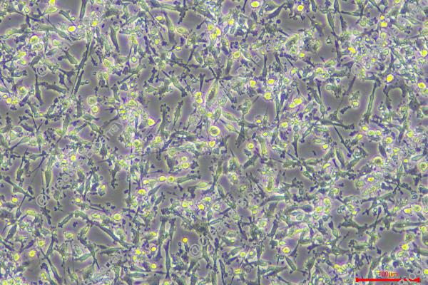
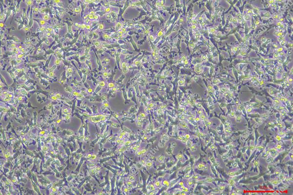
CAL-62
Cat.No.: CSC-C0480
Species: Homo sapiens (Human)
Source: Thyroid Gland
Morphology: epithelial-like cells growing in monolayers
Culture Properties: monolayer
- Specification
- Background
- Scientific Data
- Q & A
- Customer Review
Immunology: cytokeratin +, cytokeratin-7 +, cytokeratin-8 +, cytokeratin-17 -, cytokeratin-18 +, cytokeratin-19 +, desmin +, endothel -, EpCAM +, GFAP -, neurofilament +,
CAL-62 is a human ATC cell line originally derived from the right lobe tumor of a 70-year-old female patient. These cells exhibit epithelial-like, adherent morphology in vitro and forms aggressive, tumorigenic xenografts in immunodeficient mice, and has become a widely used experimental model for the biology of aggressive thyroid cancer. The line has a relatively short population doubling time and exhibits cytogenetic complexity as expected for a high-grade tumor. Key oncogenic alterations were identified in this line during genomic profiling, most notably a KRAS p.G12R mutation, as well as additional alterations in genes such as EP300 and CREBBP.
Phenotypically, CAL-62 has been shown to often lack classical markers of thyroid differentiation (e.g. thyroglobulin) and exhibit mesenchymal-like, migratory features typical of dedifferentiated ATC cells. Functionally, it has been used to study pathways involved in proliferation, invasion, metabolic reprogramming, and resistance to therapy, including radioresistance (reported in one of the earliest characterization studies of this line). Because this cell line is extensively profiled in genomic, transcriptomic, proteomic, and pharmacologic public datasets, it is an ideal model for mechanistic studies and preclinical testing of candidate therapeutics.
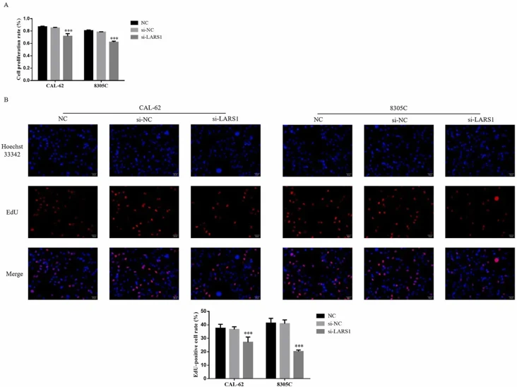
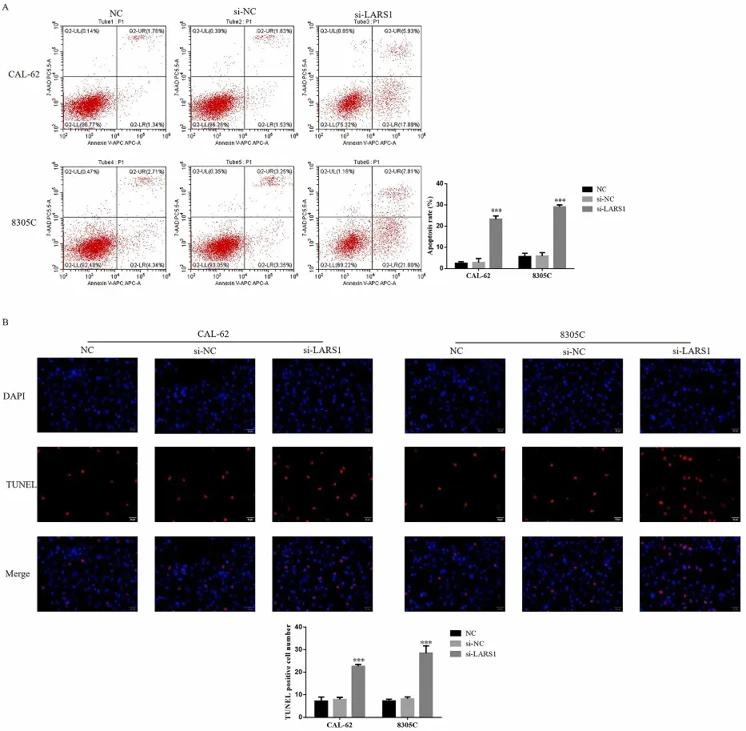
LARS1 Knockdown Decreases the Proliferation and Increases the Apoptosis of TC Cells
Given the elevated prevalence of thyroid cancer in China and its status as the leading malignant endocrine tumor, identifying molecular targets for intervention is critical. Jin's team sought to define the functional significance of LARS1 in thyroid cancer pathogenesis and delineate the mechanistic pathways involved.
Immunohistochemical analysis showed that LARS1 protein levels were much higher in thyroid carcinoma (TC) tissues than in adjacent non-cancerous tissues. This suggests that elevated LARS1 expression may contribute to TC development and progression. CCK-8 analysis revealed significantly reduced growth in LARS1-silenced CAL-62 and 8305C cells compared to controls (Fig. 1A). EdU incorporation analysis also showed a significant decrease in replicating cells after LARS1 knockdown (Fig. 1B). Flow cytometry showed a significant increase in apoptotic cells in LARS1-silenced CAL-62 and 8305C lines compared to controls (Fig. 2A). TUNEL assays confirmed this, with a significant rise in DNA fragmentation-positive cells in the si-LARS1 group compared to controls (Fig. 2B).
Ask a Question
Write your own review
- You May Also Need
Description: established from the thyroid gland from a 66-year-old male patient with thyroid gland anaplastic carcinoma

Description: Established in 1996 from the tumor tissue of a 57-year-old Caucasian man with local recurrence of a previously iodine-irradiated follicular thyroid cancer (well-differentiated with dispersed poorly ...

Description: Established from the sternal metastasis of follicular thyroid carcinoma removed from a 70-year-old Chinese woman in 1993; described to carry IGF-1 receptors

Description: established from the lymph node metastasis of a 63-year-old woman with anaplastic papillary thyroid carinoma; cells were described to not produce hormones, but to be partly positive for ...

Description: Established from cancer cells disseminated in the pleural fluid of a 44-year-old woman with undifferentiated giant cell carcinoma of the thyroid after chemotherapy, x-ray hyperthermia and OK432 ...
- Adipose Tissue-Derived Stem Cells
- Human Neurons
- Mouse Probe
- Whole Chromosome Painting Probes
- Hepatic Cells
- Renal Cells
- In Vitro ADME Kits
- Tissue Microarray
- Tissue Blocks
- Tissue Sections
- FFPE Cell Pellet
- Probe
- Centromere Probes
- Telomere Probes
- Satellite Enumeration Probes
- Subtelomere Specific Probes
- Bacterial Probes
- ISH/FISH Probes
- Exosome Isolation Kit
- Human Adult Stem Cells
- Mouse Stem Cells
- iPSCs
- Mouse Embryonic Stem Cells
- iPSC Differentiation Kits
- Mesenchymal Stem Cells
- Immortalized Human Cells
- Immortalized Murine Cells
- Cell Immortalization Kit
- Adipose Cells
- Cardiac Cells
- Dermal Cells
- Epidermal Cells
- Peripheral Blood Mononuclear Cells
- Umbilical Cord Cells
- Monkey Primary Cells
- Mouse Primary Cells
- Breast Tumor Cells
- Colorectal Tumor Cells
- Esophageal Tumor Cells
- Lung Tumor Cells
- Leukemia/Lymphoma/Myeloma Cells
- Ovarian Tumor Cells
- Pancreatic Tumor Cells
- Mouse Tumor Cells