Studies on Prostate Tumor Cells Triggered by Photodynamic Therapy
Photochemical & Photobiological Sciences. 2023 Jun; 22 (6): 1341-1356.
Authors: de Melo Gomes LC, de Oliveira Cunha AB, Peixoto LFF, Zanon RG, Botelho FV, Silva MJB, Pinto-Fochi ME, Góes RM, de Paoli F, Ribeiro DL.
INTRODUCTION
Prostate cancer is the most common cancer in American men, aside from skin cancer. Photodynamic Therapy (PDT) has merged as a treatment to cause cell death only a century ago. Methylene blue (MB) is a thiazine dye that presents excellent photochemical properties, being a potent photosensitizer for PDT.
METHODS
- Human prostate tumor epithelial cell line-PC3 was used in this investigation and maintained in DMEM medium supplemented with 10% fetal bovine serum (FBS) and 1% penicillin/streptomycin and kept in an incubator at 37ºC and 5% CO2. We use methylene blue (MB) as a photosensitizer.
- PC3 cell viability was evaluated after treatments using an MTT-based cell titer 96 Aqueous Non-Radioactive Cell Proliferation Assay kit. PC3 cell ability to migrate under the appropriate treatments was evaluated using a cell scratch/wound healing assay.
- The immunocytochemistry assay evaluated the Bcl-2 expression (apoptosis resistance-related) and LC3 (autophagy marker). According to the instructions, active caspase-3 quantification and apoptosis/necrosis detection were performed in PC3 cells.
- Acidic vesicle quantification was used to obtain data on autophagy. Total antioxidant activity was evaluated by ferric reduction (Fe3+/Fe2+) at low pH. The malondialdehyde (MDA) level was estimated using the double heating method of Draper and Hadley. Quantification of reduced glutathione with trichloroacetic acid (TCA).
- Browse our recommendations
Creative Bioarray provides professional products and services, including but not limited to the following.
| Product/Service Types | Description |
| Cell Viability Assays | Cell viability analyses are crucial for the evaluation of cell health and compound toxicity. Creative Bioarray is capable of providing various options for cell viability assays and compound profiling services. |
| Cell Migration and Invasion Assay Services | Migration is a key property of live cells and critical for normal development, immune response, and disease processes such as cancer metastasis and inflammation. We provide cell migration and Invasion Assay Services to all of our customers. |
| Cell Apoptosis Assays | The process of programmed cell death, or apoptosis, is generally characterized by distinct morphological characteristics and energy-dependent biochemical mechanisms. We offer Cell Apoptosis Assays to all of our customers. |
RESULTS
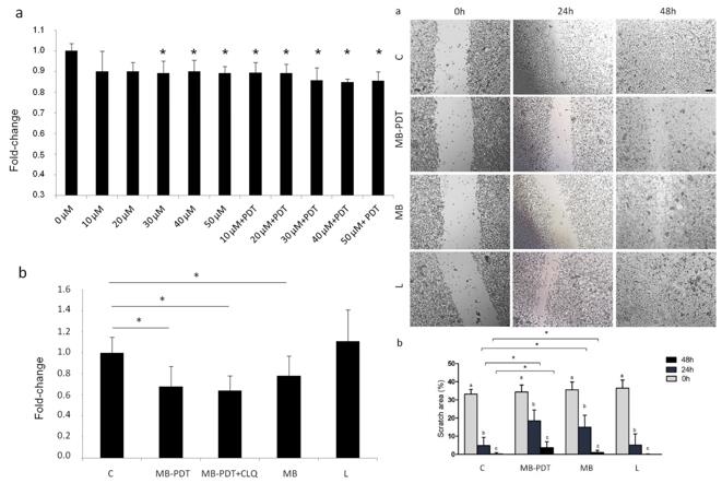
- PC3 cell viability was assessed by MTT assay. PC3 cell migratory activity was decreased after 24 and 48 h for both treatments (MB and MB-PDT) compared with the control group.
 Fig. 1 Left: Phototoxic effect of MB-PDT in PC3 cells; Right: MB-PDT reduced Cell Migration in PC3 cells.
- MB-PDT did not alter PC3 cells' apoptotic activity. According to AO quantification and LC3 puncta immunofluorescence, such findings suggested that autophagy could be triggered by MB-PDT in these cancer cells.
- Another cell death mechanism, necroptosis, was analyzed by quantifying MLKL protein content in PC3 after MB-PDT treatments. It is also involved in MB-PDT-induced cell death. Adaptive response to oxidative stress was analyzed by intracellular levels of antioxidant enzymes, total antioxidant activity, and products generated by oxidative stress. MB-PDT group is not exclusive to the combined treatment but may potentiate oxidative damage.
SUMMARY
In conclusion, MB-PDT treatment reduced the antioxidant potential and increased lipid peroxidation, affecting cell viability and migration in PC3 cells. Such an effect was associated with high autophagic activity as a pro-survival response to oxidative damage.
RELATED PRODUCTS & SERVICES
Reference
- de Melo Gomes LC, et al. (2023). "Photodynamic therapy reduces cell viability, migration and triggers necroptosis in prostate tumor cells." Photochem Photobiol Sci. 22 (6), 1341-1356.

