Simultaneous Double IF Protocol
GUIDELINE
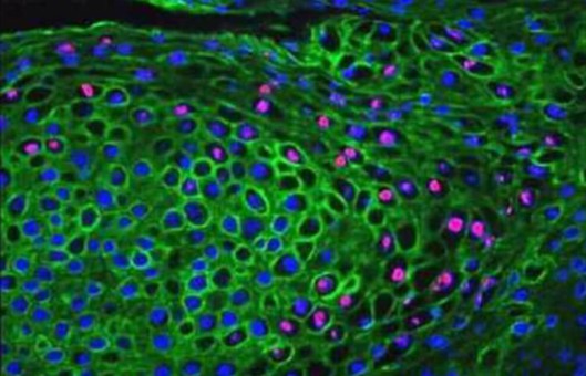
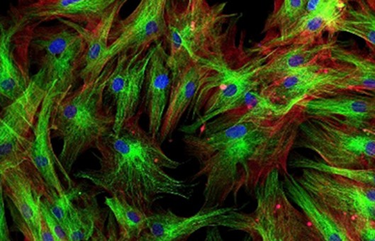
In order to be able to examine the co-distribution of two (or more) different antigens in the same sample, a double immunofluorescence procedure can be carried out. Primary antibodies raised in different species can be used either in parallel (in a mixture) or in a sequential way. We provide a protocol for immunofluorescent double staining incubating the antibodies together.
METHODS
Preparation of Slides and Samples
- Cell lines, cytology smears, cytospin preparations. Coat coverslips with polyethylineimine or poly-L-lysine for 1 hr at room temperature. Rinse coverslips well with sterile H2O (3 times 5 min each). Allow coverslips to dry completely and sterilize them under UV light for at least 4 hr. Grow cells on glass coverslips or prepare cytospin or smear preparation. Rinse briefly in phosphate-buffered saline (PBS).
- Frozen (cryostat) sections. Snap frozen fresh tissue in liquid nitrogen or isopentane pre-cooled in liquid nitrogen. Store frozen blocks at -80°C. Cut 4-8 μm thick cryostat sections and mount on super frost or gelatin coated slides. You can store slides at -80°C until needed. Before IF staining, warm up slides at room temperature for 30 minutes.
- Paraffin-embedded sections. Deparaffinize sections in xylene 2 x 5 min. Hydrate with 100% ethanol 2 x 3 min. Hydrate with 95% ethanol 1 min. Rinse in distilled water and then follow procedure for fixation and antigen retrieval as required.
Fixation
- Fix the samples either in ice-cold methanol, acetone (1-10 min) or in 3-4% paraformaldehyde in PBS pH 7.4 for 15 min at room temperature.
- Wash the samples twice with ice cold PBS.
Permeabilization
- Incubate the samples for 10 min with PBS containing 0.25% Triton X-100 (or 100 μM digitonin or 0.5% saponin). Triton X-100 is the most popular detergent for improving the penetration of the antibody. However, it is not appropriate for the use of membrane-associated antigens since it destroys membranes.
- Wash cells in PBS three times for 5 min.
Blocking and Simultaneous Incubation
- Incubate cells with 1% BSA in PBST for 30 min to block unspecific binding of the antibodies.
- Incubate cells in the mixture of two primary antibodies (e.g. rabbit against human target-1 and mouse against human target-2, if the targets are human proteins) in 1% BSA in PBST in a humidified chamber for 1 hr at room temperature or overnight at 4°C.
- Decant the mixture solution and wash the cells three times in PBS, 5 min each wash.
- Incubate cells with the mixture of two secondary antibodies which are raised in different species (with two different fluorochromes, i.e. Texas Red-conjugated against rabbit and FITC-conjugated against mouse) in 1% BSA for 1 hr at room temperature in dark.
- Decant the mixture of the secondary antibody solution and wash three times with PBS for 5 min each in dark.
Counter Staining
- Incubate cells on 0.1-1 μg/ml Hoechst or DAPI (DNA stain) for 1 min.
- Rinse with PBS.
Mounting
- Mount coverslip with a drop of mounting medium.
- Seal coverslip with nail polish to prevent drying and movement under microscope.
- Store in dark at -20°C or 4°C.
NOTES
If the target protein is expressed intracellularly, it is very important to permeabilize the cells. Acetone fixed samples do not require permeabilization.
RELATED PRODUCTS & SERVICES
For research use only. Not for any other purpose.
Resources
- FAQ
- Protocol
- Cell Culture Guide
- Technical Bulletins
-
Explore & Learn
-
Cell Biology
- Monocytes vs. Macrophages
- How to Detect and Remove Endotoxins in Biologics?
- Comparison of Different Methods to Measure Cell Viability
- What Are Myeloid Cell Markers?
- Contamination of Cell Cultures & Treatment
- Generation and Applications of Neural Stem Cells
- Stem Cell Markers
- Quantification of Cytokines
- T Cell Activation and Expansion
- Comparison of the MSCs from Different Sources
- How to Isolate and Analyze Tumor-Infiltrating Leukocytes?
- Guidelines for Cell Banking to Ensure the Safety of Biologics
- Cell Cryopreservation Techniques and Practices
- CFU Assay for Hematopoietic Cell
- Techniques for Cell Separation
- Circulating Tumor Cells as Cancer Biomarkers in the Clinic
- Biomarkers and Signaling Pathways in Tumor Stem Cells
- How to Start Your Culture: Thawing Frozen Cells
- Critical Quality Attributes and Assays for Induced Pluripotent Stem Cells
- What Is Cell Proliferation and How to Analyze It?
- Direct vs. Indirect Cell-Based ELISA
- STR Profiling—The ID Card of Cell Line
- Comparison of Several Techniques for the Detection of Apoptotic Cells
- How to Assess the Migratory and Invasive Capacity of Cells?
- What Cell Lines Are Commonly Used in Biopharmaceutical Production?
- Mesenchymal Stem Cells: A Comprehensive Exploration
- What are the Differences Between M1 and M2 Macrophages?
- Cryopreservation of Cells Step by Step
- What are PBMCs?
- Enrichment, Isolation and Characterization of Circulating Tumor Cells (CTCs)
- Neural Differentiation from Induced Pluripotent Stem Cells
- Isolation, Expansion, and Analysis of Natural Killer Cells
- Tumor Stem Cells: Identification, Isolation and Therapeutic Interventions
- Multi-Differentiation of Peripheral Blood Mononuclear Cells
- Tips For Cell Cryopreservation
- How to Decide Between 2D and 3D Cell Cultures?
- Organoid Differentiation from Induced Pluripotent Stem Cells
- Human Primary Cells: Definition, Assay, Applications
- What are Mesothelial Cells?
- How to Scale Up Single-Cell Clones?
- T Cell, NK Cell Differentiation from Induced Pluripotent Stem Cells
- Major Problems Caused by the Use of Uncharacterized Cell Lines
- IL-12 Family Cytokines and Their Immune Functions
- Cell Culture Medium
- How to Eliminate Mycoplasma From Cell Cultures?
- CHO Cell Line Development
- Troubleshooting Cell Culture Contamination: A Comprehensive Guide
- Strategies for Enrichment of Circulating Tumor Cells (CTCs)
- Unveiling the Molecular Secrets of Adipogenesis in MSCs
- How to Handle Mycoplasma in Cell Culture?
- How to Isolate PBMCs from Whole Blood?
- Spheroid vs. Organoid: Choosing the Right 3D Model for Your Research
- From Collection to Cure: How ACT Works in Cancer Immunotherapy
- Cell-Based High-Throughput Screening Techniques
- From Blur to Clarity: Solving Resolution Limits in Live Cell Imaging
- Overview of Cell Apoptosis Assays
- Live Cell Imaging: Unveiling the Dynamic World of Cellular Processes
- Optimization Strategies of Cell-Based Assays
- Live Cell Imaging: Unveiling the Dynamic World of Cellular Processes
- Cell Immortalization Step by Step
- Adherent and Suspension Cell Culture
- Cell Viability, Proliferation and Cytotoxicity Assays
- What Are CAR T Cells?
- From Blur to Clarity: Solving Resolution Limits in Live Cell Imaging
- Immunogenicity Testing: ELISA and MSD Assays
- Cultivated Meat: What to Know?
- Eosinophils vs. Basophils vs. Neutrophils
- Key Techniques in Primary, Immortalized and Stable Cell Line Development
- From Primary to Immortalized: Navigating Key Cell Lines in Biomedical Research
- Optimization Strategies of Cell-Based Assays
- 3D-Cell Model in Cell-Based Assay
- What Are the Pros and Cons of Adoptive Cell Therapy?
- 3D-Cell Model in Cell-Based Assay
- How to Maximize Efficiency in Cell-Based High-Throughput Screening?
- What are White Blood Cells?
- Immunogenicity Testing: ELISA and MSD Assays
- Types of Cell Therapy for Cancer
- Role of Cell-Based Assays in Drug Discovery and Development
- A Complete Guide to Immortalized Cancer Cell Lines in Cancer Research
- Exploring Cell Dynamics: Migration, Invasion, Adhesion, Angiogenesis, and EMT Assays
- Mastering Cell Culture and Cryopreservation: Key Strategies for Optimal Cell Viability and Stability
- Understanding Immunogenicity Assays: A Comprehensive Guide
-
Histology
- Fluorescent Nuclear Staining Dyes
- Stains Used in Histology
- Troubleshooting in Fluorescent Staining
- Immunohistochemistry Controls
- Overview of the FFPE Cell Pellet Product Lines
- Tips for Choosing the Right Protease Inhibitor
- Comparison of Membrane Stains vs. Cell Surface Stains
- Microscope Platforms
- How to Apply NGS Technologies to FFPE Tissues?
- Overview of Common Tracking Labels for MSCs
- Multiple Animal Tissue Arrays
- Cell and Tissue Fixation
- Immunohistochemistry Troubleshooting
- Cell Lysates: Composition, Properties, and Preparation
- Guides for Live Cell Imaging Dyes
- Instructions for Tumour Tissue Collection, Storage and Dissociation
- Mitochondrial Staining
- How to Choose the Right Antibody for Immunohistochemistry (IHC)
- How to Begin with Multiplex Immunohistochemistry (mIHC)
- Common Immunohistochemistry Stains and Their Role in Cancer Diagnosis
- Histological Staining Techniques: From Traditional Chemical Staining to Immunohistochemistry
- How Immunohistochemistry Makes the Invisible Brain Visible?
- Multiplexing Immunohistochemistry
- Modern Histological Techniques
- What You Must Know About Neuroscience IHC?
- Comparing IHC, ICC, and IF: Which One Fits Your Research?
- From Specimen to Slide: Core Methods in Histological Practice
- Serum vs. Plasma
-
Exosome
- Emerging Technologies and Methodologies for Exosome Research
- How to characterize exosomes?
- Classification, Isolation Techniques and Characterization of Exosomes
- Common Techniques for Exosome Nucleic Acid Extraction
- How Important are Lipids in Exosome Composition and Biogenesis?
- Collection of Exosome Samples and Precautions
- Current Research Status of Milk Exosomes
- How do PELN Deliver Drugs?
- What are the Functions of Exosomal Proteins?
- Exosome Size Measurement
- Production of Exosomes: Human Cell Lines and Cultivation Modes
- Techniques for Exosome Quantification
- Exosomes as Emerging Biomarker Tools for Diseases
- How to Apply Exosomes in Clinical?
- How to Perform Targeted Modification of Exosomes?
- Exosome Transfection for Altering Biomolecular Delivery
- Summary of Approaches for Loading Cargo into Exosomes
- Exosome Antibodies
- Exosome Quality Control: How to Do It?
- How to Enhancement Exosome Production?
- Applications of MSC-EVs in Immune Regulation and Regeneration
- The Role of Exosomes in Cancer
- What's the Potential of PELN in Disease Treatment?
- How to Efficiently Utilize MSC Exosomes for Disease Treatment?
- How to Label Exosomes?
- Unraveling Biogenesis and Composition of Exosomes
-
ISH/FISH
- ISH probe labeling method
- Multiple Approaches to Karyotyping
- RNAscope ISH Technology
- What are the Differences between FISH, aCGH, and NGS?
- CARD-FISH: Illuminating Microbial Diversity
- Comprehensive Comparison of IHC, CISH, and FISH Techniques
- In Situ Hybridization Probes
- Overview of Common FISH Techniques
- Guidelines for the Design of FISH Probes
- Small RNA Detection by ISH Methods
- Differences Between DNA and RNA Probes
- Overview of Oligo-FISH Technology
- Comparative Genomic Hybridization and Its Applications
- How to Use FISH in Hematologic Neoplasms?
- What Is the Use of FISH in Solid Tumors?
- FISH Tips and Troubleshooting
- Multiple Options for Proving Monoclonality
- FISH Techniques for Biofilm Detection
- Whole Chromosome Painting Probes for FISH
- Telomere Length Measurement Methods
- What Types of Multicolor FISH Probe Sets Are Available?
- Different Types of FISH Probes for Oncology Research
- Reagents Used in FISH Experiments
- Mapping of Transgenes by FISH
- What are Single, Dual, and Multiplex ISH?
- ImmunoFISH: Integrates FISH and IL for Dual Detection
- 9 ISH Tips You Can't Ignore
-
Toxicokinetics & Pharmacokinetics
- Toxicokinetics vs. Pharmacokinetics
- How to Improve Drug Plasma Stability?
- How Is the Cytotoxicity of Drugs Determined?
- How to Improve the Pharmacokinetic Properties of Peptides?
- Key Considerations in Toxicokinetic
- Organoids in Drug Discovery: Revolutionizing Therapeutic Research
- What Are Metabolism-Mediated Drug-Drug Interactions?
- Key Factors Influencing Brain Distribution of Drugs
- The Rise of In Vitro Testing in Drug Development
- Effects of Cytochrome P450 Metabolism on Drug Interactions
- Predictive Modeling of Metabolic Drug Toxicity
- Overview of In Vitro Permeability Assays
- How to Improve Drug Distribution in the Brain
- Organ-on-a-Chip Systems for Drug Screening
- Experimental Methods for Identifying Drug-Drug Interactions
- Traditional vs. Novel Drug Delivery Methods
- What factors influence drug distribution?
- How to Design and Synthesize Antibody Drug Conjugates?
- What Is the Role of the Blood-Brain Barrier in Drug Delivery?
- Parameters of Pharmacokinetics: Absorption, Distribution, Metabolism, Excretion
- Physical and Chemical Properties of Drugs and Calculations
- How to Conduct a Bioavailability Assessment?
- Comparison of MDCK-MDR1 and Caco-2 Cell-Based Permeability Assays
- Unraveling the Role of hERG Channels in Drug Safety
- Methods of Parallel Artificial Membrane Permeability Assays
- Pharmacokinetics Considerations for Antibody Drug Conjugates
- What are the Pharmacokinetic Properties of the Antisense Oligonucleotides?
- What Are Compartment Models in Pharmacokinetics?
- Pharmacokinetics of Therapeutic Peptides
- The Essentials of Quantitative Bioanalysis
- The 8 Costliest Mistakes in Preclinical CYP Phenotyping
- How Are Biomarkers Validated in Drug Development?
- Biomarkers vs. Functional Assays: Closing the Preclinical Gap
- When Should You Introduce ADME Tox Testing in Drug Development?
- 6 Easy Steps to Get Your In Vitro ADME Done
- From Cells to Systems: Modern Approaches to Disease Modeling
- How Can You Optimize Drug Toxicity Assessment?
- How to Choose the Right In Vitro ADME Assays for Small-Molecule Drugs
- What Are Biomarkers in Drug Discovery?
- The Bioanalysis Masterclass: Labile Metabolites
- How Genotoxicity Testing Guides Safer Drug Development
- Top 5 Pitfalls in In Vitro ADME Assays and How to Avoid Them
- 2D vs 3D Cell Culture Models: Which Is Best for Drug Toxicity Testing?
- Bioanalysis Errors: How to Spot and Fix Them Early
- Troubleshooting Common Issues in Drug Toxicity Testing
- What Is Genotoxicity in Pharmacology? Mechanisms and Sources
- Preclinical Workflow for Drug Toxicity Testing
- In Vitro ADME vs In Vivo ADME
- A Complete Guide to CYP Reaction Phenotyping in 2026
- How to Interpret CYP Phenotyping Data
- Reaction Phenotyping vs. Metabolic Stability
- The 8 Types of Drug Toxicity Every Researcher Must Know
- Why Cardiotoxicity Matters in R&D?
- What Are the Best Methods to Test Cardiotoxicity?
-
Disease Models
- Animal Models of Neurodegenerative Diseases
- Disease Models of Diabetes Mellitus
- Summary of Advantages and Limitations of Different Oncology Animal Models
- What Human Disease Models Are Available for Drug Development?
- Overview of Cardiovascular Disease Models in Drug Discovery
- Why Use PDX Models for Cancer Research?
- Preclinical Models of Acute Liver Failure
- Why Oncology Organoids Fail? How to Build Models That Work
- Organ-on-a-Chip: Is Your Microfluidic Setup Ready for Preclinical Trials?
- Oncology Model Strategy: From Screening to Validation
- How to Select the Right Preclinical Model for Drug Development
-
Cell Biology
- Life Science Articles
- Download Center
- Trending Newsletter