Overview of Human Disease Models
Nature Reviews Bioengineering. 2023 May; 11: 1-15.
Authors: Loewa A, Feng JJ, Hedtrich S.
INTRODUCTION
Human disease models can help unravel disease mechanisms, including for infectious and genetic diseases and cancer. Using appropriate human disease models in the drug development process and clinical decision-making improves the rate of clinical translation, reduces costs, and directly benefits patients. Different disease models are available covering a broad range of physiological and pathological conditions.
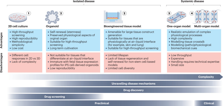 Fig. 1 Overview of different disease models.
2D Cell Cultures
- Two-dimensional (2D) cultivated patient-derived cells are an invaluable tool for studying disease phenotypes and path mechanisms, especially during the early phases of drug development.
- Primary cells are the preferred option owing to their higher genetic heterogeneity compared to cell lines, but their limited availability or in vitro proliferation capacity restricts their use. Multipotent adult stem cells (ASCs) and induced pluripotent stem (iPS) cells can help overcome this shortage, as they can readily and indefinitely propagate and convert into any somatic cell. Engineered cells, such as reporter cell lines, are also commonly used as they are amenable to high-throughput manufacturing and have high reproducibility and lower costs relative to stem cell-based methods.
Bioengineered Tissue Models
- Bioengineered tissue models are primarily generated from human stem cells or primary cells, the latter of which are isolated from surgically excised human tissue or non-transplantable organs.
- The cells are manually added or 3D printed onto or into hydrogel or polymer-based scaffolds, often in trans-well setups. Alternatively, decellularized extracellular matrix scaffolds from animal-derived organs or non-transplantable human organs can be reseeded with human cells.
- This approach is often used for multi-layer or stratified tissues such as the gut, lungs, and skin but also the brain and liver as it allows a controlled build-up of the model.
Organoids
- Organoids are self-organizing 3D structures generated from tissue-specific ASCs or iPS cells. Stem cells are embedded in an extracellular matrix that provides the scaffold for tissue growth. Organoid formation is then guided by a cocktail of growth factors that are pivotal for tissue development in vivo, which enables stem cells to maintain their differentiation and self-renewal capacity.
- Protocols for the generation of iPS cell-derived organoids are available for a variety of human organs, including the gut, stomach, liver, pancreas, lung, thyroid, kidney, retinal and optic cup, and brain.
Organs-On-Chips
- Inter-tissue crosstalk and complex in vivo-like processes cannot be mimicked in single and static tissue models. To enable tissue perfusion and dynamics as well as multi-organ crosstalk, OoCs have been introduced. OoCs are perfused microfluidic platforms that contain bioengineered or miniaturized tissues or organs interconnected by 3D microchannels to simulate the in vivo functions, biomechanics, and physiological responses of organs.
- OoCs can be single-organ or multi-organ systems. Single-organ systems can assess the response of a specific tissue or organ to a particular stimulus. By contrast, multi-organ systems allow the study of the communication and interactions of several tissues and organs simultaneously.
Creative Bioarray Relevant Recommendations
Creative Bioarray has established multiple animal disease models including cardiovascular disease models, chronic liver disease models, metabolic disease models, neurological disease models, oncology models, etc. We provide validated and custom in vivo disease modeling and services.
RELATED PRODUCTS & SERVICES

Reference
- Loewa A, et al. (2023). "Human disease models in drug development." Nat Rev Bioeng. 11, 1-15.