GUIDELINE
- Mixed lymphocyte culture (MLC) or mixed lymphocyte reaction (MLR) is a mixture of two unrelated individual lymphocytes cultured together, due to the different histocompatibility antigens on the lymphocyte membranes of the two, they can stimulate each other, resulting in the division of each other's lymphocytes to proliferate and transform. Transformed lymphoblastoid cells show increased cell size, increased synthesis of DNA in the nucleus and RNA in the cytoplasm, etc. The percentage of transformed lymphocytes can be counted morphologically or determined by measuring the amount of 3H-labeled thymidine riboside (3H-TdR) incorporation into activated lymphocytes as a precursor for DNA synthesis. The isotope labeling method is more objective, reproducible, and has more accurate results. The intensity of the proliferative reaction is proportional to the degree of difference between the two histocompatibility antigens. The worse the compatibility between the two, the stronger the reaction.
- This method has two-way and one-way MLC, in two-way MLC, both sides of the lymphocytes stimulate each other to proliferate and transform, that is, both sides of the lymphocytes are both stimulating cells and reactive cells. In one-way MLC, one side of the lymphocytes will be irradiated with X-ray or processed with mitomycin C, so that it loses the ability to proliferate and react but still retains its antigenic stimulating effect, at this time, MLC is only one side of the lymphocytes. In this case, only one side of lymphocytes in the MLC undergoes a proliferative response, so it is possible to understand the intensity of stimulation and proliferative response of lymphocytes in different individuals.
METHODS
- The lymphocyte suspensions of two individuals A and B are labeled separately, with the one of individual A as the response cell of MLC and the one of individual B as the stimulus cell of MLC, and labeled as Bm.
- Take 0.9 mL of lymphocyte suspension from individual B, add 400 μg/mL of mitomycin C 0.1 mL to make the final concentration of 40 μg/mL cell suspension, place in a 37°C water bath for 30 min, centrifuge, and discard the supernatant, the precipitated cells are washed once with culture medium, and the concentration of the cells is adjusted to 1×10⁶/mL.
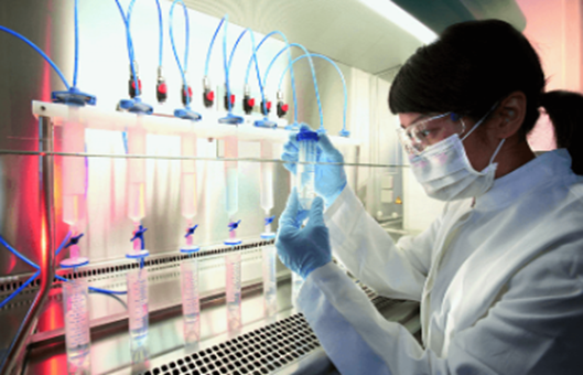
- Take 0.1 mL of reactive cells (A) and stimulated cells (Bm) in the wells of a round-bottomed cell culture plate (A Bm) with a micro-sampler, meanwhile, set up its control group AA or BBm and a blank control group. The cells are incubated at 37°C in a 5% CO2 incubator for 5 d.
- 15-16 h before the termination of culture, 3H-TdR 1 μL (containing 0.5 μCi) is added to each well using an isotope micro-sampler. Blank control group, no isotope is added at this time.
- Cells are collected on glass fiber filter paper with a cell collector, washed with saline to remove free 3H-TdR, dried in an oven at 60°C, cooled, and then placed into a measuring cup containing scintillation solution, and the intensity of radiation is detected with a α-body scintillometer. The blank control is added with an isotope before sample treatment, and the treatment method is the same as that of the experimental group.
- Expressed as counts per minute (cpm). All samples should be subtracted from the cpm value of the blank control group before data processing. The intensity of the MLC response is usually expressed in terms of the cpm value or stimulation index (SI) of the sample. It can also be expressed in terms of the net cpm value.
NOTES
- The concentration of stimulating cells and responding cells is generally 1:1 or 2:1.
- The culture time is generally 5-6 d, and the 3H-TdR doping time is generally 15-16 h before termination of culture.
- A cell culture plate with a round bottom is preferred.
- Aseptic operation should be observed during the experiment.
- This test can be used for tissue and organ transplantation mating, monitoring indicators of post-transplantation acute rejection and typing of HLA and H-2 class II antigens, and other studies.
- The positive reaction is obvious when the test is done with lymphocytes from two unrelated individuals.
RELATED PRODUCTS & SERVICES