Microscopy Studies of Synapses in Alzheimer's Disease Tissue
Seminars in Cell & Development Biology. 2023 Apr; 139: 13-23.
Authors: Griffiths J, Grant SGN.
INTRODUCTION
- Synapse loss and damage are central features of Alzheimer's disease (AD) and contribute to the onset and progression of its behavioral and physiological features. The term "synapse pathology" encompasses a multitude of pathological changes at the synapse. These include synaptic loss, dysfunction, and toxic accumulation or aggregation of proteins within the synapse.
- The study summarizes observations of synapse pathology in AD made using electron microscopy (EM), diffraction-limited light microscopy, and super-resolution nanos copy (SRN), including abnormalities in synapse density, altered morphology of dendritic spines, changes in synaptic protein levels and synaptic targets, and altered synaptic mitochondrial number.
Electron Microscopy Studies
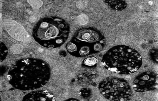
- EM approaches have been at the forefront of studies showing a correlation between synapse loss and cognitive decline. In cases with mild cognitive impairment (MCI) – a presumed prodromal AD transitional state, synapse loss was first observed in the CA1 regions of the hippocampus, inferior temporal cortex, and posterior cingulate cortex. In late-stage AD, synapse loss was further observed in the outer molecular layer of the dentate gyrus and the precuneus cortex.
- Focused ion beam milling and scanning EM (FIB/SEM) has also been used to reveal region- and layer-specific changes in dendritic spine morphology in the trans entorhinal cortex, entorhinal cortex, and the CA1 region of the hippocampal formation. In the entorhinal cortex, layer II spines were described as being larger and more complex, whereas layer III spines were smaller and "simple" when compared with controls.
Diffraction-Limited Light Microscopy Studies
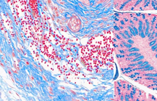
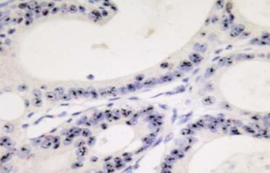
- Although EM has regularly been used to study synapse morphology in post-mortem human AD brains, it lacks the molecular specificity and throughput that can be achieved by light microscopy. Light microscopy has been used with a range of tissue staining methods, including the classical Golgi method and antibody staining. Recently, Golgi-Cox staining and 3D modeling have been used to assess dendritic spine structure in AD.
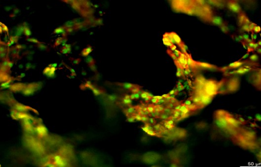
- To go beyond the axial resolution of light microscopy (~70 nm), ultra-thin tissue sectioning and deconvolution methods have been used in array tomography and fluorescence deconvolution tomography. Combined with fluorescent antibody labeling of inhibitory and excitatory synaptic proteins, these methods have been used to study synapse numbers and their association with plaque location, oligomeric Aβ, genotype, and the ratio of excitatory and inhibitory synapses in AD post-mortem brain tissue.
Super-Resolution Nanoscopy Studies
- In contrast to conventional fluorescence light microscopy, which is constrained by light diffraction limitations, SRN can resolve the subsynaptic localization of labeled proteins, revealing their differential distribution within and between synapses.
- In APP/PS1 mice, stimulated emission depletion (STED) nanoscopy revealed changes in hippocampal spine head and neck morphology. The morphology of a whole human amyloid plaque was reconstructed using STED and array tomography. STED has also been used to resolve tau filament structures in AD cortical grey matter and to validate potentially novel synaptic biomarkers from CSF in preclinical AD.
RELATED PRODUCTS & SERVICES


Reference
- Griffiths J and Grant SGN. (2023). "Synapse pathology in Alzheimer's disease." Semin Cell Dev Biol. 139, 13-23.