ISH Protocol for Electron Microscopy
GUIDELINE
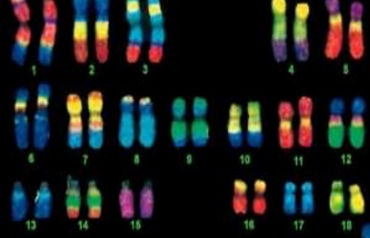
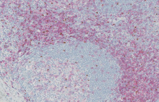
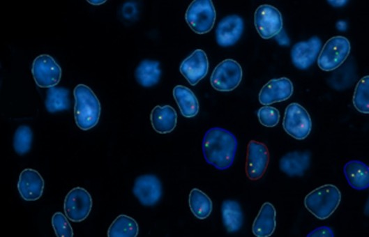
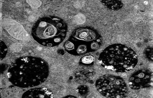
In the great majority of cases in situ hybridization is used to localize mRNA species at the tissue level, or DNA at the chromosome level. These approaches are generally best done by light microscopy. There are instances, however, when it becomes important to localize nucleic acids at the subcellular level, this brings us into the domain of the electron microscope. The distribution of nucleic acids within the cell can be an important component of their function.
METHODS
Preparing hapten-labeled RNA probes
- Linearize the template using a restriction endonuclease that does not generate a 3' overhang. If a 3' overhang cannot be avoided, blunt the ends using T4 polymerase.
- Determine the DNA concentration of the template. Prior to in vitro transcription DNA should be dissolved in DEPC-treated distilled water or TE.
- At room temperature add the following reagents in this order, 4 μL of 5× transcription buffer, 2 μL of 100 mM DTT, 1 μL of RNasin (20 U), 1 μL of 10 mM ATP, 1 μL of 10 mM GTP, 1 μL of 10 mM CTP, 0.5 μL of 10 mM UTP, 2.5 μL of 2.5 mM biotin-11-UTP (or 2.5 mM DIG-11-UTP), template DNA (0.5-2.0 μg), 1 μL of RNA polymerase (20 U). The total reaction volume should be adjusted to 20 μL with the addition of DEPC water.
- Mix the components and then incubate at 30°C for 3-4 h.
- Stop the reaction by adding 1 μL of 20% SDS.
- Set aside 3 μL for agarose gel electrophoresis.
- Add DEPC-treated water to the reaction products to bring the volume up to 50 μL.
- Pass this solution through a 30-column according to the manufacturer’s instructions to remove unincorporated nucleotides by exclusion chromatography.
- Estimate the volume of the probe solution and add 1/10 vol of 4 M LiCl, and then 2.5 vol of ethanol.
- Allow standing in a -20°C freezer for at least 1 h.
- Centrifuge in a microcentrifuge at 4°C for 15 min at 12,000g.
- Pour off the supernatant and invert the tubes to let drain for 15 min at room temperature.
- Resuspend the pellet in 10-1000 μL of hybridization buffer. The amount of hybridization buffer used will depend on the efficiency of the transcription reaction. The final probe concentration should be approx. 2-10 μg/mL.
Hybridization
- Dispense 3 μL aliquots of probe solution onto the bottom of a sterile 4 cm polycarbonate Petri dish.
- Without pretreatment of the sections, place the grids, section side down, on the probe droplets. Prehybridization was not found to be beneficial.
- Place the Petri dish into the sealable container along with a small amount of hybridization buffer (0.25 mL) to humidify the container. Seal the container.
- Hybridize at the chosen temperature for at least 1 h.
- After hybridization, grids are washed by dipping in 4×SSC, then 2×SSC, and then 1×SSC.
Probe detection
- Direct method. After blocking, incubate the grids in droplets of the anti-hapten antibody gold-conjugate diluted in SC buffer containing 1% BSA. The dilution ratio will be indicated by the product manufacturer and may have to be refined empirically. Generally, a ratio of 1:20 is appropriate. Incubation should be at room temperature and proceed for at least 60 min. Longer incubations, up to overnight, may be used to saturate probe detection depending on the size of the gold grains. For longer incubations ensure grids are in a vapor-tight container humidified with SC buffer to stop the droplets from evaporating.
- Indirect method. Following blocking, incubate in anti-hapten antibody (5 μg/mL) in SC buffer containing 1% BSA at room temperature for 30 min. Rinse grids under a gentle stream of SC buffer from a wash bottle. Incubate in secondary antibody conjugated to gold as for the direct method. After incubation with the gold-conjugate, rinse grids with SC buffer, and then with distilled water. Allow grids to air dry. Stain with uranyl acetate and lead citrate as required, remembering that excessive staining may obscure the gold labeling.
Creative Bioarray Relevant Recommendations
- ISH is a technique that allows for precise localization of a specific segment of nucleic acid within a histologic section. We provide a wide range of ISH products and services, including ISH Probes, ISH Reagents, In Situ Hybridization (ISH) & RNAscope Service, Digital ISH Image Quantification and Statistical Analysis, and others.
NOTES
- RNA is easily destroyed by ribonucleases which are extremely resilient enzymes and common contaminants from our skin and untreated solutions. Hence, it is important to wear gloves while preparing probes and to use DEPC-treated water and buffers.
- Both DIG and biotin are effective hapten probe labels and should both produce reliable results. The option of haptens is primarily useful when considering double labeling for distinct target molecules where the different hapten types can be used for the different probes.
RELATED PRODUCTS & SERVICES
Reference
- Asp J, et al. (2006). "Nonradioactive in situ hybridization on frozen sections and whole mounts." Methods Mol Biol. 326, 89-102.
