Brain Organoid Development from hPSCs
World Journal of Stem Cells. 2023 Jun 26; 15 (6): 530-547.
Authors: Yan YW, Qian ES, Woodard LE, Bejoy J.
INTRODUCTION
- Human pluripotent stem cells (hPSCs) include both human embryonic stem cells (hESCs) and human-induced PSCs (hiPSCs). Current in vitro disease models that use hiPSCs begin with skin or blood cells that have been reprogrammed with the four transcriptional elements octamer-binding transcription factor 4, SRY-box 2, Krüppel-like factor 4, and MYC. Through differentiation, these hPSCs are the starting material to create models for different organs including the brain, kidney, liver, lung, and pancreas.
- Protocols for producing brain organoids were derived from the EB and SFEBq methods for neural induction. The formation of brain organoids is based on the self-organization and self-renewal of stem cells to generate a mixed cell population in a 3D suspension culture. Brain spheroids and organoids have been widely used to model human brain development and neurological diseases in vitro and have provided a promising platform for drug screening.
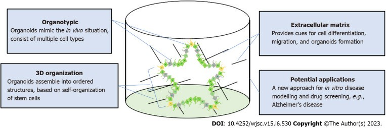
Fig. 1 Self-organization of brain organoids.
Forebrain Organoids
- The areas of the brain originating from the telencephalon and diencephalon are referred to as the forebrain. The telencephalic region comprises the cerebral cortex and cerebellum, whereas the diencephalon includes the thalamus, hypothalamus, and pituitary glands. Self-organized cortical organoids or dorsal forebrain organoids were first reported using the SFEBq method. Multiple cortical layer tissues were generated by inhibiting TGF-β and Wnt signaling, resulting in dorsal-ventral patterning.
- The technology of brain organoids has also been utilized to generate organoids that can model other regions of forebrain tissue including thalamic organoids, hypothalamic organoids, and hippocampal organoids. Hippocampal spheroids can be derived from hiPSCs using dual SMAD inhibition, Shh, and Wnt pathway inhibition followed by Wnt activation. Commonly reported hippocampal markers include zinc finger and BTB domain-containing and Prospero homeobox 1. Hippocampal spheroids can be used to model AD pathology either by the exogenous addition of amyloid beta oligomer or by using amyloid precursor protein/PS1 variant hiPSCs.
Midbrain Organoids
- The protocol to differentiate human midbrain-like organoids (hMLOs) employs several molecules to mediate the differentiation of neuroepithelial cells. These factors include hBDNF, hGDNF, dibutyryl cyclic adenosine monophosphate, ascorbic acid, TGF-β3, and purmorphamine.
- To scale up the generation of midbrain organoids, one study recently published microfabricated disk technology using eNUVIO EB-Disks. Another study found that the use of recombinant spider-silk microfibers functionalized with full-length human laminin produced similar ventral midbrain organoids with lower inter-organoid variability. Alternatively, an automated approach, termed automated midbrain organoids, was published that produced high-throughput 3D midbrain organoids. The high-throughput production of hMLOs from hPSCs in spinner flasks was also reported using TH-TdTomato reporter hPSC lines as well.
Hindbrain Organoids
- The medulla, pons, and cerebellum make up the hindbrain region, which is developed from the metencephalon and myelencephalon. Methods to generate hindbrain organoids commonly involve purmorphamine-mediated Shh signaling activation to convert neuroepithelial cells into ventral identity neurons. RA is used as a potent caudalizing agent to promote the fate of hindbrain cells instead of Wnt signaling activation, which is required for midbrain patterning.
- The first published granule cell differentiation protocol using hiPSCs involved several factors including FGF8B, Wnt proteins, BMPs, and RA. This method recapitulates anteroposterior and dorsoventral patterning and thereby induces MATH1-expressing mitotic neural progenitors, which can later be differentiated into cerebellar granule cells.
- Recently, it has been published a protocol that generates mature cerebellar neurons without the need for such a co-culture system. This method stimulates the development of cerebellar precursors with FGF19, followed by self-organization and differentiation using stromal cell-derived factor 1 and BDNF/GDNF.
Creative Bioarray Relevant Recommendations
Creative Bioarray developed a protocol that can differentiate brain organoids from iPSCs. We also provide a characterization of iPSC-derived organoids using immunocytochemistry and multi-electrode array (MEA).
RELATED PRODUCTS & SERVICES
Reference
- Yan YW, et al. (2023). "Neural lineage differentiation of human pluripotent stem cells: Advances in disease modeling." World J Stem Cells. 15 (6), 530-547.