GUIDELINE
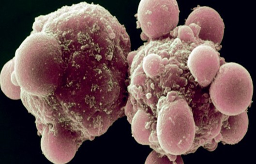
- Apoptosis or programmed cell death (PCD) is one of the cellular life phenomena, which was proposed by Kerr in 1972. It plays a very important role in embryonic development, tissue repair, and clearance of self-reactive T lymphocytes in the body. The deregulation of apoptosis can lead to the development of various clinical diseases. For example, Alzheimer's disease is associated with excessive apoptosis of neural cells, and autoimmune diseases and tumors are associated with inhibition of apoptosis. Apoptosis is distinct from necrosis in that it has a series of cytomorphological and biochemical changes, including the appearance of chromatin condensation, DNA degradation, and apoptotic vesicle formation.
- In apoptosis, a large number of sticky 3'-OH ends are generated by double-strand breaks or single-strand breaks of chromosomal DNA, which can be labeled to the 3'-ends of DNA by deoxyribonucleotide end transferase (TdT) and derivatives formed by luciferin, peroxidase, alkaline phosphatase or biotin, allowing the detection of apoptotic cells. These methods are called terminal-deoxynucleotidyl transferase-mediated nick end labeling (TUNEL). Since normal or proliferating cells have little DNA breakage and thus no 3'-OH formation, they can rarely be stained. This makes TUNEL the most common method for detecting DNA fragmentation (apoptosis).
METHODS
Detection of apoptosis by flow cytometry (AnnexinV-FITC/PI double-staining method)
- The cell suspensions are collected in 15 mL centrifuge tubes, centrifuged at 2000 r/min for 5 min, and the supernatant is discarded. After digestion with 0.25% Trypsin, add an equal volume of cell base culture medium to terminate the reaction, collect the cell suspension into a 15 mL centrifuge tube, centrifuge at 2000 r/min for 5 min, and discard the supernatant.
- Resuspend the cells with 1× Binding Buffer for 1×10⁶/mL.
- Take 100 μL of the above suspension into a flow tube.
- Add 5 μL Annexin V-eGFP to the cell suspension, mix gently, and incubate for 15 min at 2-8°C under light-proof conditions.
- Add 10 μL of 100 μg/mL PI and mix gently and incubate for 5 min at 2-8°C under light-proof conditions.
- Add 400 μL of Binding Buffer and mix well for 1 h before performing the flow-through assay.
TUNEL kit for apoptosis detection
- Prepared cell crawls are fixed with 4% PFA for 30 min at room temperature and washed 3 times with PBS.
- The membrane-breaking solution is broken 2 times for 5min each and washed 3 times for 2min each with PBS.
- Cover the cells with 50 μL of equilibration buffer. Place at room temperature for 5-10min to equilibrate.
- Blot off most of the 50 μL of equilibration buffer with blotting paper around the equilibrated area, then add 50 μL of rTdT incubation buffer (pre-configured on ice, protected from light). Wet box and incubate for 60min at 37°C protected from light.
- Add 300 μL/well 2×SSC and leave it at room temperature for 15 min to terminate the reaction.
- Immerse cell crawls in PBS and leaves at room temperature for 5min. Repeat washing 3 times to remove unadulterated fluorescein-12-deoxyuridine triphosphate and remove non-specific staining.
- Stain with 1×hoechst for 15min and wash 3 times with PBS.
- Observe the staining under a fluorescent microscope and take pictures (Red and green light are optional as needed).
Creative Bioarray Relevant Recommendations
- Creative Bioarray offers a wide range of Cell Apoptosis Assay Kits, including Annexin V Apoptosis Kit, Caspase Assay Kit, DNA Fragmentation Assay Kit, and Mitochondrial Apoptosis Detection Kit.
NOTES
- Be gentle during the operation and do not blow the cells with force.
- Perform analysis as soon as possible after the experimental manipulation is completed to avoid quenching of fluorescence with time.
- Double staining requires flow cytometry to adjust fluorescence compensation, so cells with various fluorescence single staining are needed as control.
- When choosing a fixative, we recommend using 4% paraformaldehyde for fixation, not ethanol and methanol, and not formaldehyde instead, because most of the commercially available formaldehyde contains methanol. This will make our subsequent staining process less efficient in labeling and make it difficult to observe fluorescence.
- The false positive experimental results may also be related to the staining incubation step.
- When performing fluorescence microscopy observation, depending on the microscope, specific parameters exposure time is recommended within 1000 ms, gain parameters are recommended within 500%, or no gain exposure time control again within 2000 ms, overexposure will also increase the false positives of the experiment.
RELATED PRODUCTS & SERVICES