GUIDELINE
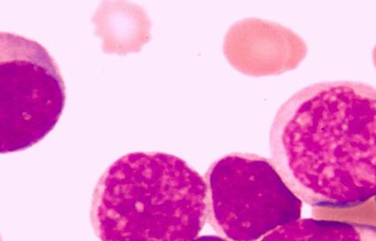
- To grasp the proliferation rate of cells, the common methods include cell count at different time points, the MTT method, and the CCK8 method to draw proliferation curves. The following is a brief description of the method for plotting proliferation curves using the CCK8 method as an example.
- The basic principle is that the reagent contains WST-8. In the presence of the electron carrier 1-methoxy-5-methylphenazinium dimethyl sulfate (1-Methoxy PMS) is reduced by the cellular dehydrogenase to a highly water-soluble yellow met san product (Formazan dye). The amount of metazoan produced is proportional to the number of living cells and inversely proportional to the cytotoxicity. For the same cells, there is a linear relationship between the shade of color and the number of cells. The number of living cells can be indirectly reflected by measuring the OD value at 450 nm using an enzyme marker.
METHODS
- Firstly, the apposed cells in the logarithmic phase are digested by adding trypsin to dislodge the cells.
- When the cells are mostly shed, add twice the volume of trypsin-containing medium to terminate the digestion and transfer the cell suspension to a centrifuge tube.
- Centrifuge at 1000r/min for 3-5min.
- Discard the supernatant of the centrifuge tube and resuspend it with 1 mL of serum-containing medium.
- Dilute the cell suspension according to the counting result to ensure the cell concentration is 2×10⁵/mL.
- Select the appropriate size plate according to the need, here take a 96-well plate as an example. Set up a blank control, 7 experimental groups, and 5 replicate wells for each group. Add 100 μL of the diluted cell suspension to each well.
- After 12-24 hours of general inoculation and good cell apposition, aspirate the medium of each well. Add medium without drug to the blank group and medium containing different concentrations of drug to the experimental group. Put into the incubator and incubate for several hours (here different drugs may have different action times, some are 4 hours, some are 12 hours, and some are 24 hours, choose according to the actual situation).
- After the drug treatment, aspirate the medium from each well again. At this point, add the medium containing CCK-8.
- Put it into the incubator for 0.5-4 hours, and take it out for testing when the color changes to orange. Note that this step should be observed at any time, the color change should not be too long.
- Take out the 96-well plate and put it into the enzyme standard for detection, the wavelength is set to 450nm.
- Plots are made with different growth times and absorbance values, with the horizontal axis being time points and the vertical axis being absorbance values.
Creative Bioarray Relevant Recommendations
- Cell proliferation assays are used to monitor the dynamic growth of a cell population or to detect daughter cells in a growing population. We provide cell analysis kits and a variety of reagents for studying cell proliferation.
NOTES
- CCK-8 can be stored for at least 6 months at 0-5°C and 1 year at -20°C protected from light.
- If the OD value is not measured temporarily, 10uL of 0.1M HCl solution or 1% (w/v) SDS solution can be added to each well and stored in a refrigerator at 4°C with the plate covered and protected from light, and the absorbance will not change when measured within 24 hours.
- If the substance to be measured is oxidizing or reducing, the drug effect can be removed by changing the fresh medium before adding CCK8. Of course, the drug influence is relatively small, you cannot change the medium, directly deduct the blank absorption after adding the drug in the medium can be.
- When incubated in the incubator, the outermost ring of the culture plate is the most prone to dryness and volatilization, increasing the error due to inaccurate volume. In general, the outermost circle of the hole with medium or PBS is not used as the assay hole.
- Before testing with the enzyme marker, you need to ensure that there are no air bubbles in each well, otherwise it will interfere with the determination.
- Phenol red and serum do not interfere with the CCK8 assay and can be eliminated by deducting the absorbance of the background in the blank wells.
- CCK8 can detect E. coli, but not yeast cells. Bacterial contamination needs to be avoided during each determination of the cell proliferation assay to avoid affecting the results.
- Metals affect CCK-8 color development. When the final concentration is 1 mM of lead chloride, ferric chloride, and copper sulfate will inhibit 5%, 15%, and 90% of the color development reaction and degrade the sensitivity. If the final concentration is 10 mM, it will inhibit 100%.
RELATED PRODUCTS & SERVICES